
Embolism
Definition/classification
Mass carried in circulation obstructing CV system = embolus. 90% = thromboemboli.
By composition: solid/liquid/gas. By infection: bland/septic. By source: cardiac/arterial/venous/lymphatic. By flow: paradoxical (venous↔arterial via PFO/septal defect/AV shunt) vs retrograde (against flow, e.g. prostate Ca→spine via valveless intraspinal veins + raised pressure).
Thromboembolism
Arterial: 80-85% intracardiac (mural thrombi LA/LV, vegetations, prosthetic valves, cardiomyopathy) + arterial sources (plaques, aneurysm, paradoxical). Effects: infarction (limb 70-75%>spleen>kidney>brain>intestine), gangrene, mycotic aneurysm, MI, sudden death.
Venous: DVT legs (most common) > pelvic > upper limb > cavernous sinus > right heart. → pulmonary embolism.
Pulmonary thromboembolism
95% from popliteal/femoral/iliac veins. Risk: stasis + hypercoagulability. Saddle embolus (bifurcation) or multiple/fragmented (lower lobes). Paradoxical embolism possible via septal defect.
Consequences (by size/number):
- Sudden death (massive) — or delayed MI-like picture
- Acute cor pulmonale (numerous small) — acute RHF + vasoconstriction
- Pulmonary infarction (small branch) — chest pain, haemoptysis, dyspnoea
- Pulmonary haemorrhage (terminal endarteries)
- Resolution (60-80% small emboli) — fibrinolysis, often silent (bronchial circulation compensates)
- Chronic sequelae (repeated, organise not resolve) — pulm HTN, chronic cor pulmonale, pulm arteriosclerosis
Fat embolism
Traumatic (long bone fracture=commonest, ortho surgery, soft tissue) vs non-traumatic (burns, DM, fatty liver, pancreatitis, sickle cell, decompression sickness, hyperlipidaemia, CPB).
Pathogenesis (combine): mechanical (trauma→marrow fat→lung) · emulsion instability (non-traumatic, chylomicron/FA aggregation) · intravascular coagulation (DIC-related aggregation) · toxic injury (FFA→vessel injury→permeability→oedema).
Pulmonary: common autopsy finding post-fracture, vacuoles on H&E, needs frozen section+Sudan/oil red O/osmic acid. Systemic (via PFO/AV shunt/vertebral plexus): brain (petechiae, microinfarcts, delirium→coma→death), kidney (↓GFR, tubular damage), skin/conjunctiva petechiae, fat in urine/sputum.
Gas embolism
Air embolism:
- Venous — neck surgery/trauma, obstetric, IV infusion, angiography. 100-150mL fatal (40mL if severely ill). Position matters (upright→brain via SVC). Death = air trapped in RV/pulmonary trunk or froth in arterioles. Confirm: open heart/PA under water.
- Arterial — cardiothoracic surgery, paradoxical (PFO), arteriography. Marble skin, retinal air bubbles, tongue pallor, sudden death (coronary/cerebral, smaller volumes suffice).
Decompression sickness (caisson disease/divers’ palsy/aeroembolism) — sudden decompression (diver ascent too fast, or unpressurised high-altitude), N2 comes out of solution (fat-affinity). Worse in obese.
- Acute: “bends” (joint pain), “chokes” (resp distress), cerebral (vertigo/coma/death)
- Chronic: avascular necrosis (femoral head/tibia/humerus), CNS ischaemia (paraesthesia/paraplegia), lung (haemorrhage/oedema/emphysema), skin, hepatic/pancreatic lipid vacuoles
Amniotic fluid embolism
Most serious/unpredictable/unpreventable maternal mortality cause. Labour/postpartum: squames, vernix, lanugo, meconium bile, mucus → uterine veins → RH (via tears or forced sinusoidal entry).
Morphology: pulm haemorrhage/congestion/oedema/ARDS + RH dilatation + identifiable amniotic contents.
Clinical: sudden resp distress, cyanosis, shock, convulsions, coma, death. Mechanisms: mechanical block + anaphylactoid reaction + DIC (amniotic thromboplastin) + haemorrhage (thrombocytopenia/afibrinogenaemia).
Atheroembolism
Eroded plaque (aortic) → cholesterol crystals/hyaline/calcified debris → medium/small arteries + foreign body reaction. → ischaemia/infarcts (kidney/spleen/brain/heart), limb gangrene, HTN (if renal).
Tumour embolism
Vascular invasion → metastasis. Classic: renal clear cell Ca, lung Ca, melanoma.
Misc emboli
Tissue/placental fragments, RBC sludging, bacteria, parasites, barium (enema), foreign bodies.
Pulmonary thrombosis vs thromboembolism
| Thrombosis (local) | Thromboembolism | |
|---|---|---|
| Origin | Locally formed | Travelled |
| Location | Small vessels | Major vessels |
| Attachment | Firm | Loose/free |
| Gross | Head pale/tail red | No head-tail distinction |
Clinical relevance
60-80% resolution + clinical silence (bronchial circulation) = why many PEs undiagnosed; minority that organise→chronic thromboembolic pulm HTN. Fat embolism theory (mechanical=trauma; emulsion instability=metabolic) helps predict clinical setting beyond fracture. Paradoxical (needs shunt) vs retrograde (needs reversed flow, no shunt) = distinct mechanisms. Amniotic fluid embolism mortality driven by DIC+anaphylactoid as much as mechanical block.
Definition and Classification
Embolism is partial or complete obstruction of the cardiovascular system by any mass carried in the circulation; the transported mass, detached from its site of origin, is an embolus. The great majority (~90%) of emboli are thromboemboli — thrombi or thrombus fragments detached from the vessel wall (see Thrombosis).
Emboli are classified several ways:
- By composition — solid (thromboemboli, atheromatous material, tumour cell clumps, tissue fragments, parasites, bacterial clumps, foreign bodies), liquid (fat, amniotic fluid, bone marrow), gaseous (air, other gases).
- By infection status — bland (sterile) or septic (infected).
- By source — cardiac (left atrium/ventricle, endocarditis vegetations), arterial (systemic arteries — brain, spleen, kidney, intestine), venous (pulmonary arteries), or (rarely) lymphatic.
- By flow behaviour — a paradoxical embolus crosses from venous to arterial circulation (or vice versa) via an abnormal communication (patent foramen ovale, septal defect, pulmonary AV shunt); a retrograde embolus travels against the normal direction of blood flow (e.g. prostate carcinoma metastasising to spine via retrograde flow through valveless intraspinal veins under raised intra-abdominal/thoracic pressure from coughing or straining).
Thromboembolism
Arterial (systemic) thromboembolism arises chiefly (80–85%) from intracardiac sources — mural thrombi in the left atrium/ventricle, valve vegetations, prosthetic valves, cardiomyopathy — or less often from arterial sources (atherosclerotic plaques, aortic aneurysm, paradoxical embolism). Effects depend on embolus size, lodgement site, and collateral adequacy: infarction (lower limb 70–75%, spleen, kidney, brain, intestine — in that order of frequency), gangrene (if collaterals are inadequate), mycotic aneurysm (from infected/septic emboli in endocarditis), myocardial infarction (coronary embolism), and sudden death (coronary or middle cerebral artery embolism).
Venous thromboembolism arises from deep vein thrombosis (DVT) of the lower legs (most common), pelvic veins, upper limb veins, cavernous sinus thrombi, or right heart thrombi; its principal consequence is pulmonary embolism.
Pulmonary Thromboembolism
The most common and most fatal form of venous thromboembolism, distinct from the uncommon entity of primary pulmonary thrombosis (which arises locally, e.g. in pulmonary atherosclerosis/hypertension, and differs on gross/microscopic examination — see comparison below).
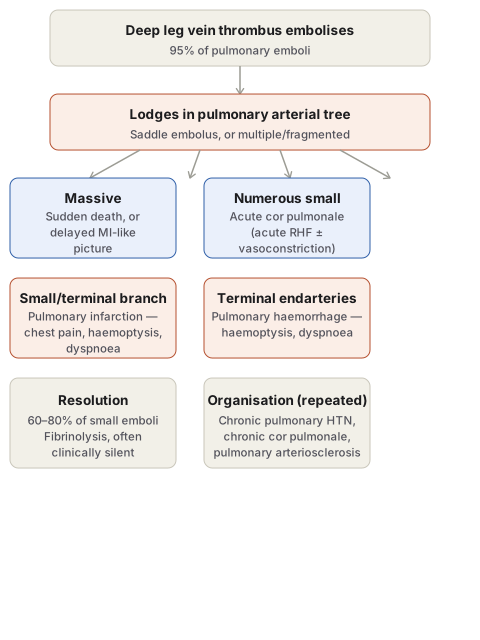
Aetiology: thrombi from the popliteal, femoral and iliac veins account for 95% of pulmonary emboli; less commonly, superficial leg varicosities or pelvic veins (periprostatic, periovarian, uterine, broad ligament). Risk factors are venous stasis and hypercoagulable states (see Thrombosis). A large thrombus may impact at the main pulmonary artery bifurcation (saddle embolus) or lodge in the right ventricle/outflow tract; more commonly, multiple or fragmented emboli lodge in several vessels, particularly the lower lobes. Rarely, a paradoxical embolus can cross a septal defect into the systemic circulation.
Consequences, depending on embolus size, number, and cardiovascular reserve:
- Sudden death — massive embolism causes instantaneous death without preceding chest pain/dyspnoea; if death is delayed, features resemble myocardial infarction (chest pain, dyspnoea, shock).
- Acute cor pulmonale — numerous small emboli obstructing most of the pulmonary circulation cause acute right heart failure, compounded by reflex/platelet-mediated pulmonary vasoconstriction.
- Pulmonary infarction — obstruction of smaller pulmonary arterial branches; chest pain (fibrinous pleuritis), haemoptysis, dyspnoea.
- Pulmonary haemorrhage — obstruction of terminal branches (endarteries) causes central haemorrhage; haemoptysis, dyspnoea, occasionally chest pain; may coexist with infarction.
- Resolution — the majority (60–80%) of small pulmonary emboli resolve completely via fibrinolysis, often clinically silent because bronchial circulation maintains parenchymal perfusion.
- Chronic sequelae — pulmonary hypertension, chronic cor pulmonale, and pulmonary arteriosclerosis follow repeated small thromboemboli that organise rather than resolve.
Fat Embolism
Obstruction of arterioles/capillaries by fat globules (fat-tissue embolism if by fragments of adipose tissue). Traumatic causes (commonest) — long bone fracture (fatty marrow enters circulation), bone concussion, orthopaedic surgery, soft-tissue laceration, pelvic fat injury in puerperium. Non-traumatic causes — extensive burns, diabetes mellitus, fatty liver, pancreatitis, sickle cell anaemia, decompression sickness, bone/soft-tissue inflammation, exogenous fat/oil introduction, hyperlipidaemia, cardiopulmonary bypass.
Pathogenesis (mechanisms may combine): the mechanical theory explains traumatic cases — fat globules released from injured bone/soft tissue enter venous circulation, are mostly arrested in pulmonary vessels, with some passing through to lodge systemically; the emulsion instability theory explains non-traumatic cases — plasma lipids (chylomicrons, fatty acids) aggregate from disturbed natural emulsification; the intravascular coagulation theory — stress-triggered DIC promotes fat globule aggregation; the toxic injury theory — high free fatty acid levels chemically injure pulmonary vessels, raising permeability and causing pulmonary oedema.
Consequences: Pulmonary fat embolism is a frequent autopsy finding after bone fracture death, usually not obstructive enough to cause symptoms from small globules alone, but widespread embolism can cause sudden death; microscopically, hyperaemia, oedema, petechial haemorrhage, ARDS-like changes, with fat appearing as vacuoles in routine stains (frozen section with Sudan/oil red O/osmic acid stains required for confirmation). Systemic fat embolism occurs when globules pass through the lung (via patent foramen ovale, pulmonary AV shunts, vertebral venous plexus) to lodge in brain (leptomeningeal/parenchymal petechiae, microinfarcts, delirium/convulsions/stupor/coma/death), kidney (glomerular capillary involvement, reduced GFR, tubular damage), and elsewhere (skin/conjunctival/serosal petechiae, fat in urine/sputum).
Gas Embolism
Air embolism occurs when air enters venous or arterial circulation. Venous air embolism — from head/neck surgery or trauma (opened neck veins), obstetric procedures (uterine venous sinus/endometrial vein entry), IV infusion under positive pressure, or venous angiography; effects depend on air volume (100–150 mL generally fatal, less in severely ill patients — as little as 40 mL), rapidity of entry, and patient position (upright position risks air ascending to the brain via the SVC); death results from air trapped in the pulmonary arterial trunk/right heart, or widespread pulmonary arteriolar occlusion by froth (confirmed at autopsy by opening the heart/pulmonary artery under water to detect escaping froth). Arterial air embolism — from cardiothoracic surgery/trauma, paradoxical passage of venous air through a PFO/pulmonary AV shunt, or arteriography; produces marble skin (cutaneous vessel blockage), visible retinal air bubbles, tongue pallor (lingual artery occlusion), and — with even small volumes — sudden death from coronary or cerebral arterial air embolism.
Decompression sickness (caisson disease, divers’ palsy, aeroembolism) results from sudden decompression — either divers/caisson workers ascending too rapidly from high atmospheric pressure, or unpressurised high-altitude flight/ascent — causing dissolved gases (chiefly nitrogen, which has particular affinity for fatty tissue) to come out of solution as bubbles that may coalesce into emboli. Severity depends on depth/altitude reached, exposure duration, rate of pressure change, and patient factors (worse in obese individuals, since nitrogen is more fat-soluble). Acute form: “the bends” (joint/ligament/tendon pain), “the chokes” (pulmonary bubble accumulation, respiratory distress), cerebral effects (vertigo, coma, death). Chronic form: ischaemic necrosis throughout the body, especially bone — avascular necrosis of femoral head, tibia, humerus; CNS ischaemic necrosis (paraesthesia, paraplegia); pulmonary haemorrhage/oedema/emphysema/atelectasis; skin changes (itching, erythema, cyanosis, oedema); hepatic/pancreatic lipid vacuolation.
Amniotic Fluid Embolism
The most serious, unpredictable and unpreventable cause of maternal mortality: during labour or the immediate postpartum period, amniotic fluid contents (epithelial squames, vernix caseosa, lanugo hair, meconium bile, mucus) enter uterine veins and reach the right heart, via myometrial/endocervical tears or forceful uterine contraction driving fluid into uterine sinusoids. Morphology: pulmonary haemorrhage, congestion, oedema, ARDS-like change, with identifiable amniotic contents in pulmonary microcirculation, plus right heart dilatation. Clinically: sudden respiratory distress, deep cyanosis, cardiovascular shock, convulsions, coma, unexpected death — from mechanical pulmonary blockage, an anaphylactoid reaction to amniotic components, DIC (thromboplastin release from amniotic fluid), and haemorrhage from thrombocytopenia/afibrinogenaemia.
Atheroembolism
Eroded atheromatous plaque (especially aortic) forms emboli of cholesterol crystals, hyaline debris and calcified material, lodging in medium/small arteries and provoking a foreign body reaction. Effects: ischaemia/atrophy/necrosis distal to occlusion; infarcts (kidney, spleen, brain, heart); lower limb gangrene; hypertension if renal vasculature is widely affected.
Tumour Embolism
Malignant cells invading local vessels form emboli that lodge elsewhere, producing metastatic deposits — notable examples: renal clear cell carcinoma, lung carcinoma, malignant melanoma (see Invasion and Metastasis).
Miscellaneous Emboli
Tissue fragments, placental fragments, red cell aggregates (sludging), bacteria, parasites, barium (post-enema), and foreign bodies (needles, talc, sutures, bullets, catheters).
Comparison: Pulmonary Thrombosis vs Thromboembolism
| Feature | Pulmonary thrombosis (local) | Pulmonary thromboembolism |
|---|---|---|
| Pathogenesis | Formed locally | Travelled from a distance |
| Location | Small arteries/branches | Major arteries/branches |
| Attachment | Firmly adherent | Loosely attached or free-lying |
| Gross | Head pale, tail red | No head/tail distinction; smooth, dry, dull surface |
| Microscopy | Platelets/fibrin in layers, lines of Zahn rare | Mixed with blood clot |
Clinical Relevance
- The 60–80% spontaneous resolution rate of small pulmonary emboli, and their clinical silence due to bronchial circulation, is the reason many pulmonary emboli go undiagnosed — it also explains why chronic thromboembolic pulmonary hypertension develops specifically from the minority that organise instead of resolving.
- Recognising which theory of fat embolism applies (mechanical in trauma, emulsion instability in non-traumatic hyperlipidaemic/diabetic states) helps anticipate the clinical setting in which fat embolism syndrome should be suspected beyond the classic long-bone-fracture scenario.
- Distinguishing paradoxical from retrograde embolism is a specific, testable distinction: paradoxical embolism requires an abnormal vascular communication (PFO, septal defect), while retrograde embolism requires only reversed flow direction under raised pressure, without any structural shunt.
- Amniotic fluid embolism’s mortality is driven as much by DIC and anaphylactoid reaction as by mechanical obstruction, which is why management extends well beyond simply treating a presumed pulmonary blockage.
Diagram 1: Consequences of Pulmonary Thromboembolism
Draw a two-stage downward column (source, then lodgement), then branch into six side-by-side outcome pairs grouped by embolus burden.
- Source: deep leg vein thrombus (95% of cases).
- Lodgement: pulmonary arterial tree — saddle embolus or multiple/fragmented.
- Branch by size/number into six outcomes, paired by mechanism: massive → sudden death; numerous small → acute cor pulmonale; small branch occlusion → pulmonary infarction; terminal endartery occlusion → pulmonary haemorrhage; most small emboli → resolution; repeated organising emboli → chronic pulmonary hypertension/cor pulmonale.
Labels required
- Each outcome box must be explicitly linked to the embolus characteristic that produces it (size, number, whether it resolves or organises) — the exam-relevant skill is predicting the consequence from the embolic burden described in a vignette, not just listing the six possible outcomes.
- The 95% figure for deep leg vein origin and the 60–80% resolution figure should both be retained as specific, frequently tested numbers.
Errors commonly made
- Treating pulmonary infarction as the default or most common outcome of pulmonary embolism, when resolution without infarction is actually the majority outcome for the small emboli that make up most cases.
- Conflating acute cor pulmonale (numerous small emboli, acute right heart strain) with chronic cor pulmonale (repeated organising emboli over time, chronic pulmonary hypertension) — these are different entities on different timescales, not the same finding at different severities.
My Sticky Notes
Personal revision notes, mnemonics and reminders.
