
Basic Diagnostic Cytology
Overview
Two branches: exfoliative (older, cells spontaneously shed from epithelium, ↑ in disease; clinician preps, pathologist interprets separately) and interventional (mainly FNAC, cytopathologist does the procedure himself).
| Feature | Exfoliative | Interventional |
|---|---|---|
| Cells | Shed naturally | Aspirated |
| Smears | Need screening to find cells | Cell-rich |
| Basis | Individual cell morphology | Cell groups+patterns |
Applications: tumour diagnosis, inflammatory/infectious conditions, cytogenetics, hormonal status (vaginal smears), cell-of-origin ID.
Nuclear criteria of malignancy
Nuclear features dominate. Key: ↑N:C ratio, irregular nuclear membrane, coarse irregular chromatin clumping+parachromatin clearing (more significant than hyperchromasia alone), irregular angular nucleoli (more significant than just more/bigger). Multinucleation and increased mitoses = unreliable alone; abnormal mitoses = significant.
Exfoliative cytology
Gynaecologic (Pap smear types)
- LVS (lateral vaginal) — hormonal assessment
- Vaginal pool/vault — endometrial/ovarian cancer detection
- Cervical (Ayre’s spatula) — cervical carcinoma
- Combined/Fast — routine screening, 97% cervical + 90% endometrial cancer detection
- Triple (CVE) — localises lesion, hard to prepare
- Endocervical/endometrial aspiration
Normal cells: superficial/intermediate/parabasal/basal squamous cells (maturity gradient). Variants: navicular cells (luteal/pregnancy/menopause), lactation cells, endocervical cells, endometrial cells (up to day 12), trophoblastic cells (post-abortion/delivery). Plus neutrophils, lymphocytes, plasma cells (chronic cervicitis), macrophages, Döderlein bacilli.
Non-gynaecologic — sample types
Respiratory (sputum, BAL/washings/brushings), GI (endoscopic lavage/brushing), Urinary (sediment, bladder washings, catheterisation, prostatic massage), Body fluids (effusions; CSF/synovial/amniotic/hydrocele/seminal/nipple discharge — small volume), Other (buccal smears for sex chromatin).
Technique
Sputum: examine 3 successive days. Effusions/CSF/semen: send unfixed, examine immediately. Usually wet-fixed (some air-dried like blood smears). Standard fixative: Papanicolaou’s (ether + 95% ethanol, equal parts). Staining: Papanicolaou/H&E (wet-fixed), Romanowsky — Leishman/MGG/Wright (air-dried).
Interventional cytology (FNAC)
Applications: palpable masses (breast, lymph nodes, thyroid, soft tissue), also salivary glands, testis, abdominal lesions; imaging-guided: prostate, pelvis, bone/joint, lung, retroperitoneum, orbit.
Advantages over surgical biopsy: OPD, no hospitalisation, usually no anaesthesia, quick/safe/minimal pain, repeatable, fast turnaround (2-24hr vs 2-4 days), cost-effective, cytopathologist gets first-hand clinical info.
Procedure: fix mass → clean skin → insert needle → suction (~10ml) + move needle back-forth → RELEASE suction BEFORE withdrawing (critical — withdrawing under suction pulls material uselessly into syringe barrel) → pressure 2-3min (prevent haematoma) → express onto slide.
Smear prep: crush-smear semisolid aspirates gently; pull fluid/blood like a PBF. Split: wet-fixed (95% ethanol, Pap/H&E, best nuclear detail) + air-dried (Romanowsky, best cytoplasm/background) — most use both.
Ancillary studies: special stains (Alcian blue/mucicarmine/PAS for mucin; methyl violet/Congo red for amyloid; bacterial/fungal stains), microbiological culture, cell block (paraffin), immunocytochemistry, image analysis/morphometry, flow cytometry, EM, molecular biology (ERBB-2, BCL-2, FISH etc).
Key site note: Abdominal fat aspiration = accepted method for secondary systemic amyloidosis — Congo red rings around fat cells, apple-green birefringence under polarised light.
Complications: haematoma (commonest, esp breast/thyroid), infection (rare even transabdominal), pneumothorax (~20% lung FNAC, usually self-resolving), tumour dissemination (theoretical systemic spread; rare needle-tract seeding in lung/prostate/pancreas cancers; ovarian cyst spillage risk).
Contraindications: coagulopathy (not thrombocytopenia alone) for deep sites; PTI <80% for liver; obstructive jaundice (bile peritonitis risk); emphysema/pulmonary HTN for lung; acute pancreatitis; acute prostatitis (transrectal, sepsis risk); acute epididymo-orchitis (defer); suspected phaeochromocytoma (BP swings).
Limitations: small sample → reliability depends on adequacy/representativeness. Negative FNAC + strong clinical suspicion → repeat FNAC or biopsy, don’t accept at face value.
Clinical relevance
Nuclear-dominant weighting of malignancy criteria is the conceptual backbone across all cytology — chromatin clumping/irregular membrane matter more than simple hyperchromasia/mitotic count, that’s real diagnostic reasoning not memorisation. Release-suction-before-withdrawal is a small technique detail with big consequence — get it backwards and an adequate sample becomes non-diagnostic purely from error. Congo red+apple-green birefringence on fat aspirate = practical minimally-invasive amyloidosis diagnosis, distinct from the more commonly taught renal-biopsy route. FNAC’s false-negative risk (from small sample size) is why a negative cytology report never overrides strong clinical suspicion — testable clinical-judgment point, not a footnote.
Overview
Diagnostic cytology is the study of individual cells (rather than tissue architecture) for diagnosis, and has two main branches: exfoliative and interventional cytology.
| Feature | Exfoliative cytology | Interventional cytology |
|---|---|---|
| Cell samples | Exfoliated from epithelial surfaces | Obtained by intervention/aspiration |
| Smears | Require screening to locate suitable cells | Abundance of cells in most smears |
| Diagnostic basis | Individual cell morphology | Cell patterns and morphology of cell groups |
| Key morphologic criteria | Nuclear characteristics most important | Nuclear characteristics important; cytoplasmic character and background equally significant |
Exfoliative cytology is the older branch — the study of cells spontaneously shed off epithelial surfaces into body cavities or fluids, aided by the fact that exfoliation rate is enhanced in disease states; cells may also be actively obtained by scraping/brushing/washing (abrasive cytology). Samples are prepared by the clinician and forwarded to the pathologist, who interprets them in isolation.
Interventional cytology is dominated by Fine Needle Aspiration Cytology (FNAC), also called Aspiration Biopsy Cytology (ABC). Unlike exfoliative cytology, the cytopathologist typically performs the procedure and interacts with the patient directly.
Applications span: diagnosis/management of benign and malignant tumours, non-neoplastic/inflammatory and infectious conditions, cytogenetics, and assessment of hormonal status in women (vaginal smears accurately reflect female sex hormone levels, e.g. confirming menopause onset) and identification of cell of origin (e.g. spermatogenic elements in testicular aspirates for male infertility workup).
Nuclear Criteria of Malignancy
Cytomorphological cancer recognition relies chiefly on nuclear characteristics; cytoplasmic features assist typing (keratinisation → squamous cell carcinoma; mucin droplets → adenocarcinoma; melanin pigment → melanoma).
| Feature | Malignant change |
|---|---|
| Nuclear size | Usually larger than benign; variation in size (anisonucleosis) more significant |
| N:C ratio | Increased |
| Nuclear shape | Moderate to marked variation |
| Nuclear membrane | Irregular thickening, angulation, indentations |
| Nuclear chromatin | Hyperchromasia (less significant); uneven distribution, coarse irregular angulated clumping, parachromatin clearing (more significant) |
| Nucleoli | Increased size/number (less significant); irregular angular outlines (more significant) |
| Number of nuclei | Multinucleation unreliable — nuclear character matters more |
| Mitoses | Increased mitoses unreliable — abnormal mitoses significant |
Exfoliative Cytology
I. Gynaecologic Exfoliative Cytology
Smears from the female genital tract are traditionally called “Pap smears.” Different preparation methods serve different purposes:
- Lateral vaginal smears (LVS) — upper third of lateral vaginal walls; ideal for cytohormonal assessment.
- Vaginal ‘pool’/‘vault’ smears — posterior fornix; recommended for detecting endometrial and ovarian cancer.
- Cervical smears — Ayre’s spatula from the cervical portio; ideal for cervical carcinoma detection.
- Combined (“Fast”) smears — vaginal pool + cervical scraping combined; recommended for routine population screening, detecting up to 97% of cervical and ~90% of endometrial cancers when properly prepared.
- Triple (cervical-vaginal-endocervical, CVE) smears — three distinct samples on separate areas of one slide; allow lesion localisation but are technically difficult to prepare.
- Endocervical/endometrial smears — via aspiration of the respective canal/cavity contents.
Normal cells in combined smears: four squamous epithelial cell types (superficial, intermediate, parabasal, basal), differing in size, nuclear character, and cytoplasmic staining — increasingly small and basophilic from superficial to basal, reflecting decreasing maturity. Notable variants: navicular cells (boat-shaped intermediate cells, seen in luteal phase/pregnancy/menopause), lactation cells (acidophilic parabasal cells during lactation), endocervical cells (columnar, honeycombed clusters), endometrial cells (seen up to day 12 of cycle), trophoblastic cells (post-abortion/delivery). Non-epithelial elements include neutrophils, lymphocytes, plasma cells (chronic cervicitis), macrophages, and Döderlein bacilli (normal vaginal flora).
II. Non-Gynaecologic Exfoliative Cytology
| Organ system | Sample types |
|---|---|
| Respiratory tract | Sputum; bronchial washings/brushing/bronchoalveolar lavage (BAL) |
| Gastrointestinal tract | Endoscopic lavage/brushing |
| Urinary tract | Urinary sediment; bladder washings; retrograde catheterisation; prostatic massage secretions |
| Body fluids (large volume) | Effusions |
| Body fluids (small volume) | CSF, synovial fluid, amniotic fluid, hydrocele fluid, seminal fluid, nipple discharge |
| Other | Buccal smears (for sex chromatin) |
Techniques of Exfoliative Cytology
- Timing/handling: for sputum, examination of specimens on three successive days is recommended; bronchoscopic material must reach the lab without delay; effusions, CSF, and semen must be sent unfixed and examined immediately.
- Fixation: material is usually wet-fixed; some smears (like blood films) are air-dried instead.
- Standard fixative: Papanicolaou’s fixative — equal parts ether and 95% ethanol.
- Processing: some samples (sputum, fluids) need laboratory preparation, including centrifugation.
- Staining: Papanicolaou and H&E stains for wet-fixed smears; Romanowsky stains (Leishman’s, May-Grünwald-Giemsa, Wright’s) for air-dried smears.
Interventional Cytology
Interventional cytology includes fine needle aspiration cytology, imprint cytology, crush smear cytology, and biopsy sediment cytology, but is virtually synonymous with FNAC.
Applications of FNAC
Most often used for palpable mass lesions: breast masses, enlarged lymph nodes, enlarged thyroid, superficial soft tissue masses; also salivary glands, palpable abdominal lesions, testicles; and (with imaging guidance) prostate, pelvic organs, bone/joint spaces, lungs, retroperitoneum, orbit.
Advantages of FNAC (vs Surgical Biopsy)
- OPD procedure, no hospitalisation required.
- No anaesthesia needed (except specific circumstances).
- Quick, safe, minimally painful.
- Can be repeated without much inconvenience.
- Rapid turnaround (2–24 hours, vs 2–4 days for histopathology).
- Cost-effective, minimal equipment.
- Cytopathologist gains first-hand clinical knowledge by performing the procedure, aiding interpretation.
General Procedure
Materials: syringe with fine needle (25–20 gauge depending on site — finer for lymph nodes/children, longer/spinal-puncture-type for lung/abdomen/prostate/ovary, coarser 18-gauge for bone), syringe holder (e.g. Franzen handle), glass slides, 95% ethyl alcohol fixative.
Method: palpate and fix the mass → clean skin → insert needle → apply suction (~10 ml) while moving needle back and forth within the lesion → release suction before withdrawing the needle (withdrawing under suction pulls material uselessly into the syringe barrel) → apply pressure to puncture site 2–3 minutes to prevent haematoma → express aspirate onto a slide using air pressure.
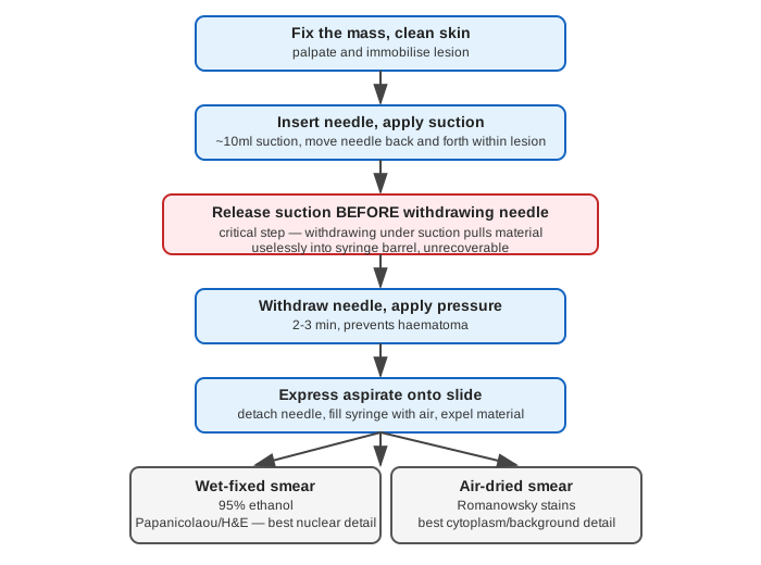
Smear preparation: semisolid aspirates are crush-smeared with a slide/coverslip under gentle even pressure; fluid/blood droplets are pulled like a peripheral blood film. Smears are split — half wet-fixed in 95% ethanol (Papanicolaou/H&E staining, best nuclear detail) and half air-dried (Romanowsky staining, best cytoplasmic/background detail); most cytopathologists use both.
Special and Ancillary Studies on Aspirates
- Special stains — Alcian blue/mucicarmine/PAS (mucin, carbohydrates); methyl violet/Congo red (amyloid); bacterial/fungal stains.
- Microbiological culture — viral, fungal, mycobacterial, bacterial.
- Cell block — paraffin-embedded centrifuged cell button, allows histologic-pattern recognition and further ancillary study.
- Immunocytochemistry — antibody panels applied to smears/cell block sections.
- Image analysis/morphometry — quantifies N:C ratio, nuclear area/shape/size.
- Flow cytometry — ploidy status, S-phase fraction.
- Ultrastructural studies — TEM/SEM.
- Molecular biology — oncogene detection (ERBB-2 in breast cancer, BCL-2 in lymphomas), DNA sequencing, gene expression profiling, FISH.
Site-Specific Notes
- Abdominal fat aspiration — the accepted method for diagnosing secondary systemic amyloidosis; amyloid shown as rings around fat cells with Congo red staining (congophilia) and apple-green birefringence under polarised light.
- Radiological guidance — non-palpable lesions require localisation (plain X-ray for bone, fluoroscopy/CT for chest/abdomen, ultrasonography as the most versatile, real-time, radiation-free option — though not useful for deep intrathoracic or bony lesions).
Complications and Hazards
- Haematoma — commonest complication (esp. breast, thyroid); minimised by firm pressure 2–3 minutes post-procedure.
- Infection — not a significant hazard even with transabdominal aspiration through bowel wall; transrectal aspiration in acute prostatitis can cause bacteraemia/septicaemia.
- Pneumothorax — in ~20% of transcutaneous lung aspirations; usually resolves spontaneously; transient haemoptysis may accompany.
- Tumour dissemination — generalised lymphatic/vascular spread is only theoretical; local needle-tract seeding is rare (reported in lung, prostate, pancreas cancers); aspiration of malignant ovarian cysts can spill contents intraperitoneally.
Precautions and Contraindications
- Bleeding disorders — thrombocytopenia alone is not a contraindication; true coagulopathies (haemophilia) contraindicate aspiration of joints, chest, abdominal viscera (superficial lesions still permissible with prolonged pressure).
- Liver — prothrombin index should be checked; FNAC inadvisable if PTI <80%; obstructive jaundice is a relative contraindication (bile peritonitis risk).
- Lung — avoid in elderly patients with emphysema (pneumothorax risk) or pulmonary hypertension (haemoptysis risk).
- Pancreas — contraindicated in acute pancreatitis (aggravates inflammation).
- Prostate — transrectal aspiration in acute prostatitis risks bacteraemia/septicaemia.
- Testis — extremely painful in acute epididymo-orchitis; defer until the acute process resolves.
- Adrenal — avoid in suspected phaeochromocytoma (risk of provoking extreme blood pressure fluctuation).
Limitations of FNAC
Only a small cell population is sampled, so reliability depends entirely on sample adequacy and representativeness. An unrepresentative sample produces a false-negative result — a negative FNAC report against strong clinical suspicion of malignancy warrants repeat FNAC or surgical biopsy, not acceptance at face value. Utility is further limited by insufficient clinical information (mass size/site/character) or missing correlative investigations (imaging, haematology, biochemistry).
Clinical Relevance
- The nuclear-versus-cytoplasmic weighting of malignancy criteria (nuclear features dominant, cytoplasmic/background features supportive) is the conceptual backbone of cytologic diagnosis across every sample type in this topic — understanding why chromatin clumping and irregular nuclear membranes outweigh simple hyperchromasia or increased mitotic count is what separates rote memorisation from actual diagnostic reasoning.
- FNAC’s core safety rule — release suction before withdrawing the needle — is a small procedural detail with an outsized consequence: getting it backwards pulls diagnostic material uselessly into the syringe barrel where it cannot be recovered, turning an adequate sample into a non-diagnostic one purely through technique error.
- Congo red staining with apple-green birefringence on abdominal fat aspirate is a genuinely practical, minimally invasive diagnostic method for secondary systemic amyloidosis, worth remembering as a concrete clinical application distinct from its more commonly taught renal-biopsy context.
- FNAC’s stated limitation (small sample size driving false-negative risk) is the direct justification for the standing rule that a negative cytology report never overrides strong clinical suspicion — this is a testable point of clinical judgment, not just a procedural footnote.
Diagram 1: FNAC Procedure Sequence
Draw a single vertical chain of six boxes, top to bottom, with the fifth box forking into two side-by-side end boxes.
- Fix the mass, clean skin.
- Insert needle, apply suction (~10ml), move needle back and forth within lesion.
- Release suction BEFORE withdrawing needle (highlight this box — it is the step most often gotten backwards).
- Withdraw needle, apply pressure 2–3 min (prevents haematoma).
- Express aspirate onto slide. 6a. Wet-fixed smear — 95% ethanol, Papanicolaou/H&E, best nuclear detail. 6b. Air-dried smear — Romanowsky stains, best cytoplasm/background detail.
Labels required
- The suction-release step must be visually distinguished (different colour/highlight) from the rest of the sequence — it is the single highest-yield procedural detail in the whole topic, and diagrams that treat it as just another box in the chain undersell its importance.
- Both final smear types must show their stain name AND their strength (nuclear detail vs cytoplasmic/background detail) — the two are not interchangeable, they are deliberately complementary.
Errors commonly made
- Reversing the order of suction release and needle withdrawal — this is the most common practical error in FNAC technique and the most testable point in this diagram.
- Treating wet-fixed and air-dried smears as alternative options rather than a routine paired practice — most cytopathologists prepare both from the same aspirate specifically because each stain reveals different information.
My Sticky Notes
Personal revision notes, mnemonics and reminders.
