
Autophagy
Definition
“Self-eating” — lysosomal digestion of the cell’s own components. Survival mechanism under nutrient deprivation: the starved cell consumes and recycles its own cytoplasm/organelles for energy. Adaptive, but can shift to signalling cell death if stress persists.
Mechanism (5 steps)
- Cytosolic sensors detect nutrient deprivation (Atg genes activated)
- Phagophore (ER-derived double membrane) sequesters cytosol + organelles
- Phagophore closes → autophagosome
- Autophagosome fuses with lysosome → autophagolysosome
- Lysosomal enzymes digest contents → amino acids/fatty acids/sugars recycled to cytosol
Biologic significance
- Adaptation to starvation — linked to tissue atrophy under sustained reduced demand.
- If starvation persists beyond what autophagy can compensate for → triggers apoptosis.
- Extensive in ischaemic injury.
- Extensive in certain myopathies.
- Defence against infection — autophagic vacuoles form around intracellular microbes.
- Cancer cells lose dependence on autophagy to survive stress — a hallmark of malignant adaptation.
Clinical relevance
Cuts across nutrient stress, ischaemia, infection, myopathy, and cancer — not confined to one disease category. Extensive autophagy on biopsy = cell under major metabolic stress, potentially en route to apoptosis if unrelieved.
Definition
Autophagy, meaning “self-eating,” is the lysosomal digestion of a cell’s own components. It functions as a survival mechanism during nutrient deprivation, allowing a starved cell to stay alive by consuming and recycling portions of its own cytoplasm and organelles to generate nutrients and energy. Autophagy is therefore fundamentally an adaptive response, though under sufficiently prolonged or severe stress it can shift from preserving the cell to signalling its death.
Mechanism
Autophagy proceeds through a defined sequence of steps, each governed by a dedicated set of autophagy genes (Atg genes) whose products carry out the process.
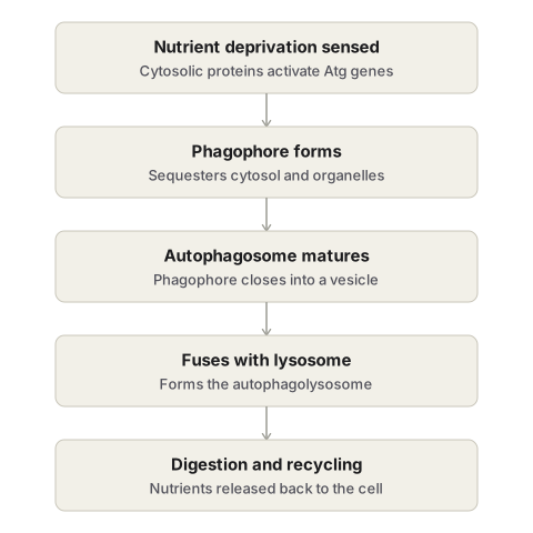
- Sensing. Cytosolic proteins detect nutrient deprivation and initiate the autophagic response.
- Sequestration. A double membrane derived from the endoplasmic reticulum, called the phagophore, begins to enclose a portion of cytosol together with intracellular organelles marked for digestion.
- Maturation of the autophagosome. The phagophore closes around its contents to form a complete double-membraned vesicle, the autophagosome.
- Fusion with the lysosome. The autophagosome fuses with a lysosome to form an autophagolysosome (also called an autolysosome).
- Degradation and recycling. Lysosomal enzymes digest the sequestered cellular components, and the breakdown products — amino acids, fatty acids, and sugars — are released back into the cytosol to be reused for the starved cell’s metabolic needs.
Biologic Significance
- Adaptation to nutrient deprivation. Autophagy is the mechanism by which a cell survives lean times by consuming its own contents, and it may be associated with the atrophy of tissue under sustained reduced demand — the two processes share this same underlying cellular strategy of downsizing to match a lower metabolic requirement.
- A route to cell death. If a starved cell can no longer sustain itself despite consuming its own contents, autophagy can itself signal a transition to apoptosis, illustrating that the boundary between an adaptive and a fatal cellular response is not fixed but depends on the severity and duration of the stress.
- Ischaemic injury. Extensive autophagy is a recognised feature of tissue subjected to ischaemia.
- Myopathies. Certain myopathies show extensive autophagic activity within affected muscle fibres.
- Defence against infection. Autophagic vacuoles can form around intracellular microbes, contributing to the destruction of these pathogens — extending autophagy’s role from a purely metabolic adaptation to a component of innate cellular defence.
- Cancer biology. Malignant cells characteristically acquire the ability to survive periods of metabolic stress without relying on autophagy, a departure from normal cell behaviour that is itself of interest in understanding tumour cell survival.
Clinical Relevance
- Autophagy sits at the intersection of several major disease processes — nutrient stress, ischaemia, infection, myopathy, and cancer — making it a mechanism worth recognising across seemingly unrelated organ systems rather than filing it under any single disease category.
- Because prolonged autophagy can tip over into apoptosis, tissue showing extensive autophagic activity on biopsy should be interpreted as a cell under significant metabolic stress, potentially on a trajectory toward cell death if the stress is not relieved.
- The loss of dependence on autophagy in cancer cells is one of several documented ways that malignant cells subvert normal cellular stress responses to gain a survival advantage, a theme that recurs across other adaptive mechanisms co-opted by tumours.
Diagram 1: Mechanism of Autophagy (five-step sequence)
Draw a single cell in cross-section, showing the sequence of vesicle formation as a series of small diagrams within it, left to right or top to bottom.
- A nucleus and cytoplasm containing normal organelles, with small dots near the membrane labelled “cytosolic sensors” to represent nutrient deprivation being detected.
- A crescent-shaped double membrane (the phagophore) beginning to wrap around a small cluster of organelles.
- The phagophore fully closed into a rounded double-membraned vesicle (the autophagosome), completely enclosing its contents.
- The autophagosome shown merging with a nearby lysosome (drawn as a smaller, denser circle), the point of fusion clearly indicated.
- The resulting single vesicle (the autophagolysosome) with its contents shown broken down into small fragments, and small arrows leaving the vesicle labelled “amino acids, fatty acids, sugars” returning to the cytosol.
Labels required
- Each of the five stages individually labelled: sensing, phagophore, autophagosome, autophagolysosome, degradation/recycling.
- The double-membraned nature of both the phagophore and the autophagosome must be drawn explicitly (two concentric lines, not one), since this is a specific ultrastructural detail examiners check.
- Atg genes/proteins should be named as the drivers of phagophore formation.
Errors commonly made
- Drawing the phagophore or autophagosome with a single membrane instead of a double membrane — this is the most common structural error on this diagram.
- Omitting the recycling step at the end, stopping the diagram at digestion without showing that the breakdown products are returned to the cell.
- Failing to distinguish the autophagosome (before fusion) from the autophagolysosome (after fusion with the lysosome) — these are two separate, individually named structures, not one.
Diagram 2: Autophagy Compared with Heterophagy
Draw two small cells side by side, each showing a vesicle being digested by a lysosome, to contrast the source of the digested material.
Left — autophagy: the vesicle being digested contains the cell’s own organelles (draw a small mitochondrion visible inside), sourced from within the same cell.
Right — heterophagy (ordinary phagocytosis): the vesicle being digested contains external material taken up from outside the cell — draw a small bacterium inside, with an arrow showing it entering from outside the cell membrane.
Labels required
- Label the source of digested material explicitly on each side: “self” for autophagy, “external” for heterophagy.
- Both pathways should be labelled as converging on the same lysosomal digestive machinery, despite differing in what is digested.
Errors commonly made
- Confusing the two terms and describing autophagy as digestion of extracellular material — the defining feature of autophagy is that the source is the cell’s own contents.
My Sticky Notes
Personal revision notes, mnemonics and reminders.
