
Autoimmune Haemolytic Anaemia
Overview
Autoantibodies against own red cells. One of 3 immunohaemolytic anaemia types (also: drug-induced, isoimmune). 2 types by thermal reactivity: warm AIHA (37°C) and cold AIHA (4°C). Coombs’ test (direct/indirect) = essential diagnostic tool.
Warm antibody AIHA
Mechanism: IgG (occ. IgA) coats red cells → splenic macrophage Fc receptors bind → strip part of membrane → acquired spherocytosis. C3 co-coating worsens haemolysis. Spleen = major destruction site.
Cause: idiopathic (most) or secondary (~25%) — SLE, CLL, lymphoma, drugs (methyldopa, penicillin).
Clinical: any age/sex, chronic anaemia with remission/relapse, splenomegaly.
Labs: mild-moderate anaemia, reticulocytosis, spherocytes on film, direct Coombs’ +ve (best at 37°C), ↑unconjugated bilirubin. Evans’ syndrome = AIHA + immune thrombocytopenia. Severe: haemoglobinaemia/uria.
Rx: remove cause, corticosteroids (1st line), transfusion if severe, splenectomy = 2nd line.
Cold antibody AIHA
Cold agglutinin disease: IgM (usually anti-I) binds RBC best at 4°C, agglutinates in cold, disaggregates on warming. Haemolysis via C3 fixation, not agglutination itself. Affects younger RBCs. Cause unknown mostly; seen with Mycoplasma, infectious mononucleosis, lymphoma.
Paroxysmal cold haemoglobinuria (PCH): IgG cold antibody (Donath-Landsteiner), anti-P antigen, complement-mediated. Cold exposure triggers attacks. Rare; assoc. tertiary syphilis, Mycoplasma, flu, measles, mumps.
Clinical (from haemolysis, not agglutination): anaemia worse in cold, Raynaud’s phenomenon, cyanosis of cold-exposed parts (nose/ears/fingers/toes), haemoglobinaemia/uria on cold exposure.
Labs: similar to warm AIHA but ↓reticulocytes (young cells hit more), LESS spherocytosis, direct Coombs’ +ve for C3 (not IgG — IgM detaches on warming).
Reference tables
RBC morphology → mechanism: spherocytes = spectrin loss (hereditary spherocytosis, AIHA) · target cells = ↑surface:volume (thalassaemia, liver disease, HbS/C) · schistocytes = trauma (microangiopathy) · sickle cells = HbS polymerisation · acanthocytes = membrane lipid abnormality (liver disease) · Heinz bodies = precipitated Hb (unstable Hb).
Extravascular vs intravascular haemolysis:
| Extravascular (common) | Intravascular (less common) | |
|---|---|---|
| Site | RE organs (spleen/marrow/liver) | Peripheral blood |
| Iron stores | ↑ | ↓ |
| Ferritin | normal/↑ | ↓ |
| Plasma Hb/methaemoglobin/Hburia/haemosiderinuria | absent | present |
| Unconj bilirubin | markedly↑ | mildly↑ |
| LDH | mildly↑ | markedly↑ |
Clinical relevance
Coombs’ test = key discriminator AIHA (+ve) vs hereditary spherocytosis (-ve) when both show spherocytes+splenomegaly+jaundice — different management (immunosuppression/splenectomy vs splenectomy alone). Warm AIHA=spleen destruction→splenectomy useful. Cold AIHA/PCH=complement-driven, partly liver clearance→splenectomy less useful. Evans’ syndrome (AIHA+ITP) = distinct combined-cytopenia pattern, changes management approach.
Overview
Autoimmune haemolytic anaemia (AIHA) results from autoantibody formation against the patient’s own red cells, one of three categories of immunohaemolytic anaemia (alongside drug-induced immunohaemolytic anaemia and isoimmune haemolytic anaemia from transfusion/pregnancy/haemolytic disease of the newborn). AIHA itself is divided by the thermal reactivity of the autoantibody into warm antibody AIHA (reactive at 37°C) and cold antibody AIHA (reactive best at 4°C). The Coombs’ (antiglobulin) test — direct (detecting antibody/complement already on the red cell) or indirect (detecting free antibody in serum) — is the essential diagnostic tool across all forms.
Warm Antibody AIHA
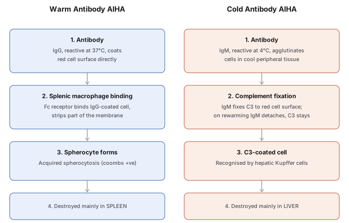
Pathogenesis: warm antibodies are usually IgG (occasionally IgA), coating the red cell surface. IgG-coated cells are bound by Fc receptors on reticuloendothelial (chiefly splenic) macrophages, which strip away part of the coated membrane — producing spherical transformation (acquired spherocytosis). Co-coating with complement (C3) promotes further red cell-leucocyte interaction and more severe haemolysis. The spleen is the major site of red cell destruction.
Aetiology: idiopathic in most cases; roughly a quarter occur secondary to an underlying immune-system disease — SLE, chronic lymphocytic leukaemia, lymphoma — or certain drugs (methyldopa, penicillin).
Clinical features: any age, either sex; chronic anaemia of variable severity with remissions and relapses; splenomegaly.
Laboratory findings: mild-to-moderate chronic anaemia; reticulocytosis; prominent spherocytosis on blood film; positive direct Coombs’ test (best detected at 37°C); positive indirect Coombs’ test if large quantities of antibody are present in serum; unconjugated hyperbilirubinaemia. Co-existing immune thrombocytopenia (± venous thrombosis) defines Evans’ syndrome. Severe cases show haemoglobinaemia/haemoglobinuria.
Treatment: remove the cause where identifiable; corticosteroids first-line; blood transfusion in severe cases; splenectomy is second-line therapy.
Cold Antibody AIHA
Occurs in two clinical settings:
1. Cold agglutinin disease — IgM antibodies, usually directed against the I antigen, bind red cells best at 4°C and agglutinate them profoundly in the cold, disaggregating rapidly on rewarming to 37°C. Haemolysis is mediated by complement (C3) fixation, not by agglutination itself. Predominantly affects younger red cells. Aetiology is unknown in most cases, but it is seen with Mycoplasma pneumoniae infection, infectious mononucleosis, and lymphoma.
2. Paroxysmal cold haemoglobinuria (PCH) — an IgG cold antibody (the Donath-Landsteiner antibody), directed against the P blood group antigen, causing complement-mediated haemolysis; attacks are precipitated by cold exposure. Uncommon; associated with tertiary syphilis, or as a complication of Mycoplasma infection, influenza, measles, or mumps.
Clinical features (from haemolysis, not agglutination itself): chronic anaemia worsened by cold exposure; Raynaud’s phenomenon; cyanosis of cold-exposed extremities (nose tip, ears, fingers, toes); haemoglobinaemia/haemoglobinuria on cold exposure.
Laboratory findings: broadly similar to warm AIHA except for thermal amplitude — chronic anaemia; low reticulocyte count (young red cells preferentially affected); less marked spherocytosis than warm AIHA; direct Coombs’ test positive for C3 on the red cell surface (not IgG, since IgM itself detaches on warming and is not detected at 37°C testing conditions).
Reference Tables (Cross-Applicable to All Haemolytic Anaemias)
Red cell morphology by mechanism: spherocytes (spectrin loss — hereditary spherocytosis, AIHA); target cells (increased surface:volume ratio — thalassaemia, liver disease, HbS/HbC disease); schistocytes (traumatic membrane damage — microangiopathy); sickle cells (HbS polymerisation); acanthocytes/spur cells (membrane lipid abnormality — severe liver disease); Heinz bodies (precipitated haemoglobin — unstable haemoglobin disorders).
Extravascular vs intravascular haemolysis: extravascular haemolysis (more common; occurs in RE organs — spleen, marrow, liver) shows increased iron stores, normal/high ferritin, absent plasma haemoglobin/methaemoglobin/haemoglobinuria/haemosiderinuria, markedly elevated unconjugated bilirubin, and only mildly elevated LDH. Intravascular haemolysis (less common; occurs directly in peripheral blood) shows the opposite pattern — decreased iron stores, low ferritin, present plasma haemoglobin/methaemoglobin/haemoglobinuria/haemosiderinuria, only mildly elevated unconjugated bilirubin, but markedly elevated LDH.
Clinical Relevance
- The Coombs’ test result is what distinguishes AIHA from hereditary spherocytosis when both present with spherocytes, splenomegaly, and jaundice — positive in AIHA, negative in the hereditary condition — directly determining whether the patient is managed with immunosuppression/splenectomy for an acquired disease or splenectomy alone for a structural one.
- Warm AIHA destruction occurring chiefly in the spleen (extravascular) versus cold AIHA/PCH’s complement-driven, partly intravascular pattern explains why splenectomy is a meaningful second-line option in warm AIHA but far less useful in cold antibody disease, where the liver (via complement receptors) is a major clearance site instead.
- Evans’ syndrome (AIHA plus immune thrombocytopenia) is a recognisable combined-cytopenia pattern worth naming specifically, since it changes the differential and management approach compared with either cytopenia occurring alone.
Diagram 1: Warm versus Cold Antibody AIHA
Draw two parallel columns (Warm, Cold), each with four stacked stages: antibody → cell interaction → resulting coated cell → site/pattern of destruction.
- Antibody — IgG at 37°C (warm) vs IgM at 4°C (cold).
- Cell interaction — Fc-receptor binding and membrane stripping (warm) vs agglutination + C3 fixation (cold).
- Resulting cell — acquired spherocyte (warm) vs C3-coated cell, IgM detached on rewarming (cold).
- Destruction site — spleen (warm) vs liver, via complement receptors (cold).
Labels required
- The antibody class and temperature must both be stated for each column — “warm” and “cold” alone are not sufficient; IgG/37°C and IgM/4°C are the tested pairing.
- Step 3 in the cold column must show that IgM itself detaches on rewarming, leaving only the C3 coating behind — this is the reason the direct Coombs’ test in cold AIHA detects C3, not IgM.
- The different destruction sites (spleen vs liver) must be explicit, since this is the mechanistic basis for splenectomy’s differing usefulness in the two conditions.
Errors commonly made
- Assuming both warm and cold AIHA produce equally prominent spherocytosis — cold AIHA spherocytosis is distinctly less marked than warm AIHA’s.
- Believing the direct Coombs’ test detects the same molecule in both conditions — it detects IgG in warm AIHA but C3 in cold AIHA, because the causative IgM antibody itself is no longer bound at testing temperature.
- Attributing cold AIHA’s haemolysis to agglutination itself rather than to complement fixation, which is the actual haemolytic mechanism.
Multiple Choice Questions (2)
My Sticky Notes
Personal revision notes, mnemonics and reminders.
