
Apoptosis
Definition
Apoptosis is programmed, internally regulated death of a single cell or a small cluster of cells, carried out by the cell’s own caspase enzymes and cleared without inflammation.
Physiological causes
Embryogenesis (interdigital web resorption) · hormone-withdrawal involution (endometrium, lactating breast) · loss of trophic stimulus (neutrophils after inflammation, lymphocytes after immune response) · deletion of self-reactive lymphocyte clones · epithelial turnover (intestine) · thymic involution
Pathological causes
DNA damage beyond repair (p53-mediated) · accumulation of misfolded protein (ER stress) · viral infection (Councilman bodies in viral hepatitis) · cytotoxic T-cell killing of tumour cells and graft cells · parenchymal atrophy after duct obstruction · low-dose injurious agents (mild hypoxia, mild radiation) · roughly 80% of cell loss in acute myocardial infarction · CD4+ T-cell depletion in HIV infection
Morphology
Light microscopy: single shrunken cell, intensely eosinophilic cytoplasm, pyknotic nucleus, no surrounding inflammation.
Electron microscopy: cell shrinkage, peripheral chromatin condensation and nuclear fragmentation, surface blebbing, formation of membrane-bound apoptotic bodies, rapid phagocytosis by macrophages.
Recognition signals for clearance
Phosphatidylserine flips from the inner to the outer membrane leaflet; thrombospondin is secreted; complement and natural antibodies may coat the apoptotic body. Together these mark the cell for prompt phagocytosis before its contents can leak out.
Mechanism
Balance between pro-apoptotic and pro-survival signals decides the outcome. Two initiating pathways converge on a common execution phase carried out by caspases (cysteine proteases cleaving after aspartate residues).
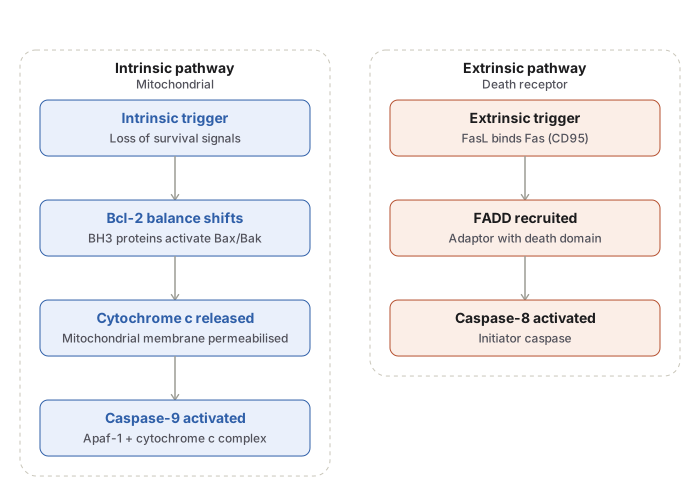
Intrinsic (mitochondrial) pathway: growth factor withdrawal, DNA damage, or ER stress activates BH3-only sensors (Bad, Bim, Bid, Puma, Noxa) → these activate Bax/Bak → mitochondrial membrane permeabilised → cytochrome c released → binds Apaf-1 → apoptosome forms → activates caspase-9. Anti-apoptotic Bcl-2, Bcl-XL, and Mcl-1 normally restrain this step.
Extrinsic (death receptor) pathway: FasL binds Fas (CD95), or TNF binds TNFR1 → FADD adaptor recruited → activates caspase-8.
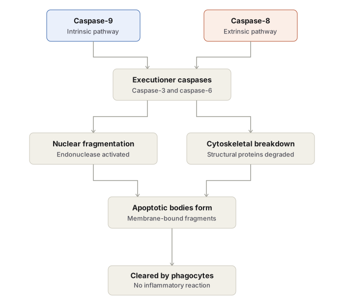
Execution phase: caspase-9 and caspase-8/10 activate executioner caspase-3 and caspase-6 → DNase activation fragments chromatin, cytoskeleton and nuclear matrix proteins degraded → cell fragments into apoptotic bodies.
Detection methods
Chromatin stains (haematoxylin, Feulgen, acridine orange) · DNA gel electrophoresis, giving a step-ladder pattern (necrosis gives a diffuse smear) · TUNEL assay · flow cytometry and propidium iodide assay · cytochrome c and caspase estimation · Annexin V staining for externalised phosphatidylserine.
Apoptosis compared with necrosis
| Apoptosis | Necrosis | |
|---|---|---|
| Cells affected | Single or small clusters | Contiguous groups |
| Cell volume | Shrinks | Swells |
| Membrane | Intact until engulfed | Ruptures early |
| Nucleus | Smooth chromatin condensation | Pyknosis, karyorrhexis, karyolysis |
| Inflammation | Absent | Present |
| Mediating enzymes | Caspases | Lysosomal hydrolases |
| DNA pattern | Step-ladder | Diffuse smear |
Disorders of dysregulated apoptosis
Reduced apoptosis — cancer, autoimmune disease. Follicular lymphoma carries t(14;18)(q32;q21), which overexpresses Bcl-2 and protects malignant B cells from apoptosis.
Increased apoptosis — neurodegenerative disease (Alzheimer, Huntington, Parkinson disease), ischaemic injury (myocardial infarction, stroke), HIV infection.
Clinical relevance
Glucocorticoids promote apoptosis of lymphoid cells; sex steroids tend to inhibit it. Loss of p53 function permits genetically damaged cells to escape apoptosis and predisposes to malignant transformation.
Definition
Apoptosis is a genetically regulated, internally programmed pathway of single-cell death, activated by the cell’s own signalling machinery rather than imposed on it from outside. The term describes a cell “falling away” from the surrounding tissue, comparable to a leaf dropping from a tree — a controlled event confined to one cell or a small cluster of cells, leaving neighbouring tissue undisturbed. It occurs both as a normal physiological process and as a mechanism of cell loss in disease, and unlike necrosis it does not release cell contents into the surrounding stroma and therefore provokes no inflammatory reaction.
Causes of Apoptosis
Physiological Situations
- Removal of surplus cells during embryogenesis, such as resorption of the interdigital webs between developing fingers and toes.
- Cyclical involution of hormone-dependent tissue on withdrawal of the driving hormone, seen in endometrial breakdown during menstruation and in regression of the lactating breast once feeding stops.
- Loss of cells after withdrawal of a trophic stimulus, as with neutrophils at the close of an acute inflammatory response, or lymphocytes once an immune response has run its course.
- Deletion of self-reactive lymphocyte clones during immune development, which prevents these cells from persisting and causing autoimmunity.
- Replacement of short-lived cells in tissues undergoing constant renewal, such as the intestinal epithelium.
- Involution of the thymus in early life.
Pathological Situations
- Elimination of cells whose DNA has been damaged beyond repair, by radiation, cytotoxic drugs, or hypoxia. The tumour-suppressor gene p53 senses such damage and, when repair is not possible, drives the cell into apoptosis; this safeguard is lost in tumours where p53 is mutated or absent, allowing genetically damaged cells to survive.
- Elimination of cells burdened by an excess of misfolded proteins. Accumulation of abnormally folded protein within the endoplasmic reticulum produces a state of ER stress that itself triggers apoptotic death, a mechanism implicated in neurodegenerative disease of the central nervous system, including Alzheimer disease, Huntington disease, and Parkinson disease.
- Death of virus-infected cells, either induced directly by the virus itself or by the host’s cytotoxic T-lymphocyte response, as in viral hepatitis, where dying infected hepatocytes form Councilman bodies.
- Death of tumour cells and of transplanted cells during graft rejection, mediated by cytotoxic T lymphocytes.
- Apoptosis of parenchymal cells following duct obstruction in glandular organs, such as the pancreas, parotid gland, or kidney, and atrophy of an organ once its trophic stimulus is withdrawn, as in prostatic atrophy following orchidectomy.
- Cell death produced by low-intensity exposure to agents that cause necrosis at higher doses — mild hypoxia, mild thermal injury, or low-dose radiation.
- A substantial share of the cell loss in acute myocardial infarction is apoptotic rather than necrotic, even though necrosis dominates the visible infarct.
- Progressive depletion of CD4+ T lymphocytes in HIV infection.
Morphology
Light Microscopy
An apoptotic cell is recognised as a single dying cell within an otherwise viable field, rather than a whole area of damaged tissue.
- The cell rounds up and shrinks into a mass with intensely eosinophilic cytoplasm.
- The nucleus shows condensed chromatin clumped as dense fragments, a change termed pyknosis.
- No inflammatory reaction accompanies the process.
Electron Microscopy
- Cell shrinkage, with dense cytoplasm.
- Nuclear condensation and fragmentation — chromatin aggregates at the margin under the nuclear membrane, and the nucleus may break into two or more fragments.
- Extensive surface blebbing, followed by fragmentation of the cell into membrane-bound apoptotic bodies, each composed of cytoplasm and tightly packed organelles, with or without a fragment of nucleus.
- Rapid phagocytosis of apoptotic cells and apoptotic bodies by macrophages, with degradation by the phagocyte’s own lysosomal enzymes; clearance is efficient enough that dying cells can disappear from the tissue within minutes, which is why extensive apoptosis rarely triggers inflammation even when many cells are involved.
Biochemical Basis of Recognition and Clearance
Apoptotic cells and apoptotic bodies undergo membrane changes that mark them out for phagocytosis before any spillage of contents can occur.
- Phosphatidylserine, ordinarily confined to the inner leaflet of the plasma membrane, flips to the outer leaflet, where it is recognised by receptors on macrophages.
- Soluble factors such as thrombospondin are secreted by the dying cell and help recruit phagocytes.
- Natural antibodies and complement proteins may coat apoptotic bodies, further assisting their removal.
Mechanism of Apoptosis
Cell survival or cell death is decided by the balance between two opposing sets of signals reaching the cell — pro-apoptotic (death) signals and pro-survival (anti-apoptotic) signals. Once the balance shifts toward death, the process unfolds through an initiation phase and an execution phase, both carried out by caspases, a family of cysteine proteases that cleave their target proteins after aspartate residues.
Initiation Phase
Two distinct pathways can initiate apoptosis, activated by different classes of stimuli.
Intrinsic (mitochondrial) pathway. This is the dominant mechanism in most physiological and pathological apoptosis, activated by signals arising within the cell. Mitochondrial permeability is governed by the Bcl-2 family of proteins — named for Bcl-2, first identified as an oncogene in B-cell lymphoma — which includes both anti-apoptotic (pro-survival) members, Bcl-2, Bcl-XL, and Mcl-1, and pro-apoptotic effector members, Bax and Bak. Growth factors and other survival signals promote production of the anti-apoptotic members; conversely, withdrawal of growth factors, DNA damage from radiation, cytotoxic drugs, or free radical injury, and accumulation of misfolded protein (ER stress) all activate a further set of Bcl-2 family sensor proteins — the BH3-only proteins, which include Bad, Bim, Bid, Puma, and Noxa. These sensors activate Bax and Bak, which form channels in the mitochondrial membrane, releasing cytochrome c and other mitochondrial proteins into the cytosol. Cytochrome c combines with a protein called Apaf-1 to form a complex called the apoptosome, which binds and activates caspase-9, the initiator caspase of this pathway. Normal cells also contain inhibitor-of-apoptosis proteins (IAPs) that hold caspase activation in check; released mitochondrial proteins neutralise these inhibitors, permitting the caspase cascade to proceed.
Extrinsic (death receptor) pathway. This pathway is triggered by signals from outside the cell, acting on death receptors — members of the tumour necrosis factor receptor family bearing a cytoplasmic “death domain.” The two principal death receptors are type 1 tumour necrosis factor receptor (TNFR1) and Fas (CD95), whose ligand is FasL. Binding of FasL to Fas recruits an adaptor protein, Fas-associated death domain (FADD), which activates caspase-8. This pathway is central to two protective functions: elimination of self-reactive lymphocytes (limiting autoimmunity) and killing of virus-infected or tumour cells by cytotoxic T lymphocytes.
Execution Phase
The two initiation pathways converge on a shared execution machinery. The mitochondrial pathway activates initiator caspase-9; the death-receptor pathway activates initiator caspase-8 (and caspase-10). These initiator caspases in turn activate executioner caspases, chiefly caspase-3 and caspase-6, which act on structural and nuclear components of the cell — activating DNase to fragment nuclear chromatin, and degrading nuclear matrix and cytoskeletal proteins to fragment the cell itself.
Methods Used to Detect Apoptosis
- Chromatin condensation demonstrated by haematoxylin and eosin, Feulgen, or acridine orange staining.
- DNA fragmentation assay by gel electrophoresis of genomic DNA, which produces a characteristic “step-ladder” pattern in apoptosis, in contrast to the diffusely smeared pattern seen when DNA is degraded during necrosis.
- The TUNEL technique, which labels fragmented DNA in situ.
- Flow cytometry, to detect rapid cell shrinkage, and propidium iodide assay.
- Estimation of cytosolic cytochrome c and of activated caspases.
- Annexin V staining, which binds phosphatidylserine exposed on the outer leaflet of the apoptotic cell membrane.
Apoptosis Compared with Necrosis
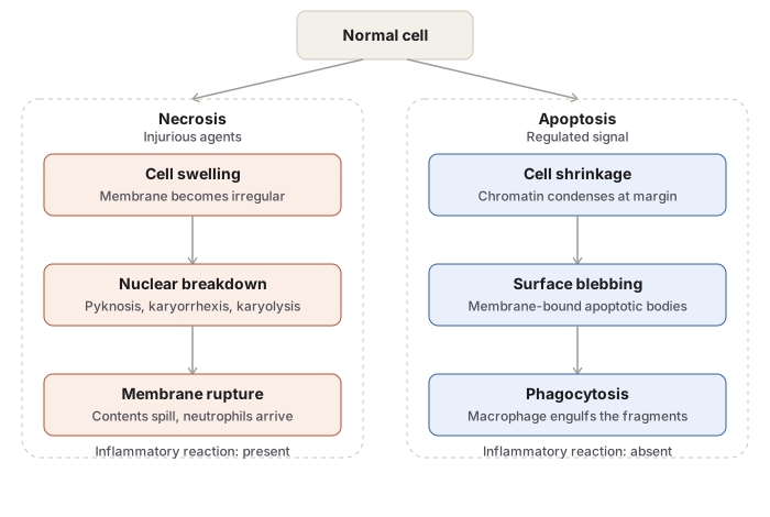
| Feature | Apoptosis | Necrosis |
|---|---|---|
| Trigger | Physiological signal, or a regulated pathological stimulus | Injurious agents — hypoxia, toxins, physical or chemical trauma |
| Cells affected | Single cells or small clusters | Contiguous groups of cells |
| Cell volume | Shrinks | Swells, then ruptures |
| Cell membrane | Intact until phagocytosed | Loses integrity early |
| Nucleus | Chromatin condenses smoothly at the margin, then fragments | Pyknosis, karyorrhexis, and karyolysis, in a disorderly sequence |
| Lysosomes | Remain intact | Rupture, releasing hydrolytic enzymes |
| Inflammatory reaction | Absent | Always present |
| Mediating enzymes | Caspases | Lysosomal hydrolases |
| DNA electrophoresis pattern | Discrete “step-ladder” bands | Diffuse smear |
Disorders Associated with Dysregulated Apoptosis
Reduced apoptosis allows abnormal or damaged cells to persist and is implicated in cancer and in autoimmune disease. The clearest example is follicular lymphoma, a B-cell non-Hodgkin lymphoma bearing the translocation t(14;18)(q32;q21), which places the Bcl-2 gene under the control of an immunoglobulin heavy-chain promoter and drives its overexpression. The resulting excess of anti-apoptotic Bcl-2 protein protects malignant B lymphocytes from apoptosis, allowing them to accumulate over long periods rather than being cleared through the normal pathway.
Increased apoptosis produces excessive loss of cells and contributes to neurodegenerative disease (Alzheimer, Huntington, and Parkinson disease), ischaemic injury in myocardial infarction and stroke, and death of virus-infected cells in conditions such as HIV infection.
Clinical Relevance
- Distinguishing apoptosis from necrosis on histology narrows the differential diagnosis: a single dying cell without surrounding inflammation points toward apoptosis, while a field of dead, inflamed tissue points toward necrosis.
- Glucocorticoids promote apoptosis in lymphoid cells, a property exploited therapeutically in lymphoid malignancy, while sex steroids tend to inhibit it.
- The Fas–FasL and mitochondrial (Bcl-2) pathways are both targets of drug development, since promoting apoptosis in tumour cells and restraining it in degenerative disease are each therapeutically desirable in their respective settings.
Diagram 1: Apoptosis and Necrosis Compared
Draw two cells side by side, each shown at three sequential stages.
Left column — apoptosis, three stages
- A normal resting cell.
- The cell shrinks, nuclear chromatin condenses into a crescent hugging the nuclear membrane, and small blebs appear on the surface.
- The cell fragments into several separate membrane-bound apoptotic bodies, each containing a portion of condensed chromatin and intact-looking organelles; draw a macrophage engulfing one fragment.
Right column — necrosis, three stages
- A normal resting cell, matching the apoptosis starting point.
- The cell swells, organelles swell, and the membrane shows irregular blebbing.
- The membrane ruptures and cell contents spill into the extracellular space; surround the ruptured cell with a scatter of neutrophils responding to the spill.
Labels required
- On the apoptosis side, label the chromatin change as pyknosis. On the necrosis side, label all three nuclear changes — pyknosis, karyorrhexis, karyolysis — at the stage each occurs.
- Label “apoptotic body” on the final apoptosis frame.
- Below each column, note the presence or absence of an inflammatory reaction.
Errors commonly made
- Drawing the apoptotic cell as swelling instead of shrinking. Shrinkage versus swelling is the single feature examiners check first when distinguishing the two processes.
- Drawing loose organelles instead of enclosed apoptotic bodies.
- Omitting the inflammatory cells around the necrotic cell.
- Labelling karyorrhexis and karyolysis on the apoptosis side, where only chromatin condensation belongs.
Diagram 2: Mechanism of Apoptosis
Draw a flowchart with one starting box branching into two parallel pathways that converge on a shared execution step.
Starting box: cell receives a death signal, branching into two arrows.
Left branch — intrinsic (mitochondrial) pathway
- Box: loss of survival signal, DNA damage, or accumulation of misfolded protein.
- Arrow to a mitochondrion drawn with a balance scale inside it — one pan labelled Bcl-2, Bcl-XL, Mcl-1 (anti-apoptotic), the other labelled Bad, Bim, Bid, Puma, Noxa (BH3-only sensors).
- Arrow to Bax/Bak forming channels in the mitochondrial membrane.
- Arrow to cytochrome c released into the cytoplasm, binding Apaf-1 to form the apoptosome.
- Arrow down to caspase-9 activated.
Right branch — extrinsic (death receptor) pathway
- Box: FasL binds Fas (CD95), or TNF binds TNFR1.
- Arrow to FADD adaptor recruited.
- Arrow down to caspase-8 activated.
Convergence point
- Both arrows merge into one box: executioner caspase-3 and caspase-6 activated.
- Arrow to a box: DNase activation fragments chromatin; nuclear matrix and cytoskeletal proteins degraded.
- Final arrow to a box: phosphatidylserine and thrombospondin exposed on the outer membrane, recognised by macrophage receptors, followed by phagocytosis.
Labels required
- Every caspase number must be written explicitly — 9, 8, 10, then 3 and 6.
- The pathway headers “intrinsic (mitochondrial)” and “extrinsic (death receptor)” must both appear.
- Fas and FasL must both be labelled.
Errors commonly made
- Assigning caspase-9 to the extrinsic pathway or caspase-8 to the intrinsic pathway.
- Drawing only one pathway and presenting it as the complete mechanism, when both converge on the same executioner caspases.
- Omitting the convergence step, leaving two disconnected pathways.
- Leaving out the final phagocytosis step.
Short Answers (1)
Multiple Choice Questions (1)
My Sticky Notes
Personal revision notes, mnemonics and reminders.
