
Amyloidosis
Definition
Extracellular deposition of insoluble fibrillar protein (amyloid) — uniform morphology/staining, heterogeneous biochemistry. Rokitansky (1842), named by Virchow (mistaken starch-like belief — iodine brown→blue with H2SO4). H&E: extracellular, homogeneous, eosinophilic hyaline.
Physical/chemical nature
Fibril proteins (95%) — non-branching, 4-6 fibrils/fibre, 7.5-10nm, cross-β-pleated sheet (→ Congo red + apple-green birefringence). = “protein misfolding disease” (proteasome/macrophage clearance fails). 2 categories: normal protein overproduced (AL, AA) vs mutant misfolding-prone (ATTR).
3 major fibril proteins:
- AL — Ig light chain (λ>κ 2:1), sequence homology present → plasma cell dyscrasias → 1° amyloidosis
- AA — 8.5kD, from 12.5kD SAA (hepatic acute-phase protein, HDL3-bound), NO sequence homology → chronic inflammation → 2° amyloidosis
- Others: ATTR (mutated/senile), Aβ2M (dialysis), Aβ (APP cleavage, Alzheimer’s), endocrine (ACal/AIAPP/AIns), APrP (prion)
Non-fibrillar (5%) — AP-component (liver, PAS+, all types, CRP-related but distinct) · apoE (apoE4→↑Alzheimer risk) · GAGs (heparan sulphate) · others.
Pathogenesis (general 4-step)
- Precursor protein pool ↑ (stimulus-driven)
- Nidus formation (BM-protein interaction, microenvironment change)
- Partial proteolysis (macrophages/RE cells) — EXCEPTIONS: ATTR (mutated, no degradation needed), Aβ2M (just accumulates unfiltered), prion (de novo β-sheet)
- Non-fibrillar stabilisation (AP/apoE/GAG) → insoluble fibril
AL-specific: plasma cell clone → excess monoclonal Ig/light chain → macrophage proteolysis → AP/GAG stabilisation AA-specific: chronic inflammation → macrophage IL-1/IL-6 → ↑hepatic SAA → RE proteolysis → AEF (amyloid enhancing factor) = nidus/accelerator → AP/GAG stabilisation
Classification
| Category | Disease | Type | Organs |
|---|---|---|---|
| Systemic 1° | Plasma cell dyscrasia | AL | Heart, bowel, skin, nerve, kidney |
| Systemic 2°/reactive | Chronic infection/inflammation/cancer | AA | Liver, spleen, kidney, adrenal |
| Haemodialysis | CRF >10yr dialysis | Aβ2M | Synovium, joint, tendon (carpal tunnel!) |
| Heredofamilial | Hereditary polyneuropathy / FMF | ATTR / AA | Nerve+heart / liver-spleen-kidney |
| Localised senile cardiac | Ageing | ATTR (unmutated) | Heart |
| Localised senile cerebral | Alzheimer’s/prion | Aβ, APrP | Brain vessels, plaques, tangles |
| Localised endocrine | Medullary thyroid Ca, T2DM | Procalcitonin, proinsulin | Thyroid, islets |
| Localised tumour-forming | — | AL | Lung, larynx, skin, bladder, tongue |
AL vs AA comparison
| AL (Primary) | AA (Secondary) | |
|---|---|---|
| Cause | Plasma cell dyscrasia (myeloma 15-20%) | Chronic infection (TB, leprosy, osteomyelitis)/autoimmune (RA, SLE, IBD)/cancer/FMF |
| Homology | Present | Absent |
| Age | >40 | Any, incl. children |
| Course | Rapid, fatal (~1yr untreated) | Better if underlying cause treated |
| Distribution | Kidney, heart, bowel, nerve | Kidney, liver, spleen, adrenal |
| Congo red after permanganate | Persists | Disappears |
Mnemonic: “3 Ps” — Persistence after Permanganate in Primary.
Heredofamilial subtypes
- ATTR hereditary polyneuropathy — AD, ~60 TTR mutations, nerve+heart, onset despite inheritance is late (past middle age)
- FMF — AR, Mediterranean populations (Sephardic Jews/Armenians/Arabs/Turks), periodic fever+polyserositis, AA type (secondary-like distribution)
- Rare: AApoAI, AGel, ALys, AFib, ACys
Staining
Iodine (historical, gross) · H&E (pink hyaline) · methyl/crystal violet (metachromasia) · Congo red + polarised light = apple-green birefringence (gold standard) · thioflavin T/S (UV fluorescence) · IHC (anti-AP=universal confirm; anti-AA/λ/κ/TTR=type-specific).
Diagnosis
Biopsy + Congo red + polarising microscopy = definitive. Best yield: renal biopsy (systemic) > rectal biopsy > abdominal fat aspirate (simple, effective) > gingiva/skin (poor). In vivo Congo red test (rare, anaphylaxis risk). Supportive: serum/urine electrophoresis, marrow aspirate.
Organ morphology
- Kidney — most common/serious (20% of amyloid deaths); glomerular BM→capillary tuft→proteinuria/nephrotic syndrome; also peritubular (casts) + arteriolar (ischaemia)
- Spleen — sago (white pulp/arterioles, modest splenomegaly, sago-grain nodules) vs lardaceous (red pulp/sinuses, marked splenomegaly ≤1kg, map-like lard areas)
- Liver — space of Disse first → hepatocyte cord compression/atrophy; function stays NORMAL even late
- Heart — pressure atrophy→restrictive cardiomyopathy, conduction system→arrhythmia; AL = ring fibres around myocardial cells; senile = LA + interatrial septum
- GI — anywhere oral-anal; tongue = tumour-forming AL → macroglossia; rectal/gingival biopsy sites
Clinical relevance
Permanganate test distinguishes AL/AA without IHC. Sago vs lardaceous spleen = classic spot diagnosis. Carpal tunnel in long-term dialysis patient → think Aβ2M amyloidosis. AL(rapid/fatal) vs AA(better if treated) prognosis difference → biochemical typing matters clinically, not just “amyloidosis” diagnosis.
Definition
Amyloidosis is a group of diseases sharing a common feature: extracellular deposition of an insoluble, fibrillar proteinaceous material called amyloid, uniform in morphological appearance, staining behaviour and physical structure across all forms, despite being biochemically heterogeneous — the precursor protein differs by clinical setting. First described by Rokitansky (1842) and named by Virchow, who mistakenly believed the material was starch-like (amylon = starch) because it stained brown with iodine and turned blue-violet with dilute sulphuric acid — a historical misnomer that has stuck despite amyloid having no relationship to starch.
By light microscopy (H&E), amyloid is extracellular, homogeneous, structureless, eosinophilic hyaline material. Different amyloid forms are named with the prefix A (for amyloid) followed by a suffix denoting the specific constituent protein — AL, AA, ATTR, and so on.
Physical and Chemical Nature
All forms of amyloid look alike morphologically but differ chemically. Two components make up the material:
I. Fibril proteins (~95% of amyloid). Electron microscopy shows a meshwork of non-branching fibrils (4–6 fibrils per amyloid fibre, each 7.5–10 nm diameter, indefinite length), each fibril itself a double helix of two β-pleated sheets. This cross-β-pleated sheet configuration (demonstrated by X-ray crystallography/infrared spectroscopy) is what gives amyloid its diagnostic staining properties — Congo red binding and apple-green birefringence under polarised light — and its alternative name, β-fibrillosis. Modern framing treats amyloidosis as fundamentally a protein-misfolding disease: normally, misfolded intracellular protein is degraded by proteasomes and misfolded extracellular protein is cleared by macrophages, but in amyloidosis this quality-control system fails, and proteins that adopt an abnormal β-pleated sheet conformation aggregate as extracellular fibrils instead of being cleared. The responsible proteins fall into two categories: normal proteins produced in excess that have an inherent tendency to self-associate (e.g. AL from excess light chain, AA from excess SAA), or mutant proteins that are structurally prone to misfolding (e.g. ATTR from a mutated transthyretin). At least 20–30 biochemically distinct proteins are now recognised to form amyloid fibrils in different clinical settings; the three most important are:
- AL (amyloid light chain) protein — derived from immunoglobulin light chain (complete or a fragment), more often λ than κ (roughly twice as common), with amino acid sequence homology between cases. Produced by immunoglobulin-secreting cells, hence associated with plasma cell dyscrasias — primary systemic amyloidosis.
- AA (amyloid-associated) protein — an 8.5 kD protein derived from a larger 12.5 kD serum precursor, SAA (serum amyloid-associated protein), an acute-phase reactant synthesised by the liver (circulating bound to HDL3) in response to chronic inflammatory/traumatic stimuli — hence elevated in chronic inflammation. Unlike AL, AA deposits show no sequence homology between cases. Underlies secondary (reactive) systemic amyloidosis.
- Other proteins, each defining its own clinicopathologic category: transthyretin (TTR) — mutated in hereditary polyneuropathic amyloidosis (ATTR), or unmutated in senile cardiac amyloidosis; β2-microglobulin (β2M) — accumulates in long-term (10–12 year) haemodialysis because it is not filtered by dialysis membranes, depositing preferentially in bone/joint/synovium; amyloid β-peptide (Aβ) — cleaved from amyloid precursor protein (APP) by β- and γ-secretase, deposited in Alzheimer disease (cerebral vessels, neurofibrillary tangles, senile plaques); endocrine hormone precursors — procalcitonin (ACal, medullary thyroid carcinoma), islet amyloid polypeptide/amylin (AIAPP, type 2 diabetes), proinsulin (AIns); and prion protein (APrP), the abnormally folded isoform underlying transmissible spongiform encephalopathies.
II. Non-fibrillar components (~5%). Chiefly amyloid P-component (AP), a liver-synthesised, PAS-positive glycoprotein present in every type of amyloid (structurally related to, but distinct from, CRP; pentagonal/doughnut profile on EM) — its universal presence is why anti-AP immunostaining confirms amyloid of any type. Also apolipoprotein E (the apoE4 allele specifically raises Alzheimer APP-deposition risk), sulphated glycosaminoglycans (notably heparan sulphate), and minor constituents (α1-antichymotrypsin, complement components, proteases). These components assist fibril stabilisation through protein aggregation and folding, protecting the deposit from resolubilisation or degradation.
Pathogenesis
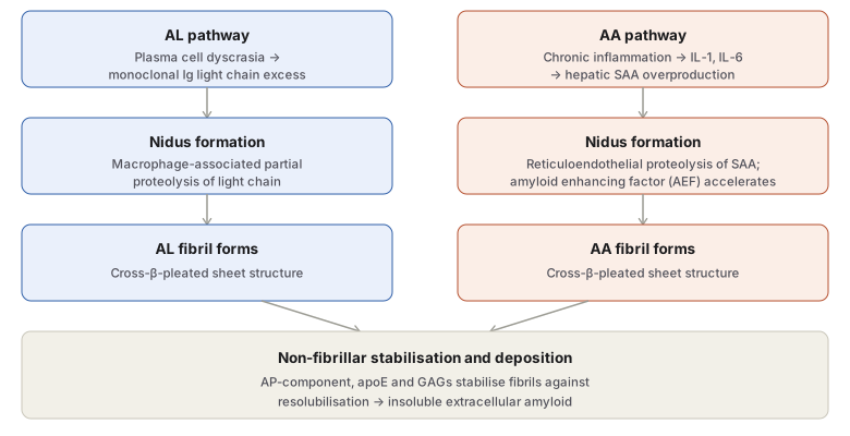
Amyloidogenesis in general follows a common sequence regardless of the specific protein involved:
- A pool of amyloidogenic precursor protein accumulates in the circulation, driven by the relevant clinical stimulus (increased hepatic AA/ATTR synthesis, increased AL synthesis by a plasma cell clone, etc.).
- A nidus for fibrillogenesis forms — an alteration in the local tissue microenvironment, involving interaction between basement membrane proteins and the amyloidogenic protein, that favours deposition.
- Partial proteolysis of the precursor typically occurs (in macrophages/reticuloendothelial cells) before fibrillar deposition — though exceptions exist: ATTR (structurally mutated, no degradation needed), β2M (simply accumulates unfiltered rather than being degraded), and prion disease (β-pleated sheet forms de novo from the normal protein).
- Non-fibrillar components (AP, apoE, GAGs) stabilise the aggregating fibrils, protecting them from resolubilisation.
AL amyloid deposition specifically: a plasma cell dyscrasia (multiple myeloma, B-cell lymphoma, other monoclonal gammopathy) drives monoclonal proliferation of plasma cells/B cells, producing excess monoclonal immunoglobulin (complete Ig, or λ/κ light chain) → partial proteolysis by closely associated macrophages → AP/GAG-assisted fibril aggregation.
AA amyloid deposition specifically: chronic inflammation (infection, autoimmune disease, malignancy) activates macrophages, which release IL-1 and IL-6 → hepatic SAA synthesis rises → partial proteolysis in reticuloendothelial cells → amyloid enhancing factor (AEF), itself produced in chronic inflammation/cancer/familial Mediterranean fever, acts as a nidus and accelerates AA fibril deposition → AP/GAG-assisted stabilisation.
Classification
| Category | Associated disease | Biochemical type | Organs typically involved |
|---|---|---|---|
| Systemic — Primary | Plasma cell dyscrasias | AL | Heart, bowel, skin, nerves, kidney |
| Systemic — Secondary (reactive) | Chronic infection/inflammation, cancer | AA | Liver, spleen, kidney, adrenals |
| Systemic — Haemodialysis-associated | Chronic renal failure (long-term dialysis) | Aβ2M | Synovium, joints, tendon sheaths |
| Systemic — Heredofamilial | Hereditary polyneuropathy / FMF / rare mutations | ATTR / AA / AApoAI, AGel, ALys, AFib, ACys | Nerves, heart / liver, spleen, kidney, adrenals |
| Localised — Senile cardiac | Ageing | ATTR (unmutated) | Heart |
| Localised — Senile cerebral | Alzheimer’s, prion disease | Aβ, APrP | Cerebral vessels, plaques, tangles |
| Localised — Endocrine | Medullary thyroid Ca, Type 2 DM | Procalcitonin, proinsulin | Thyroid, islets |
| Localised — Tumour-forming | — | AL | Lung, larynx, skin, bladder, tongue, eye |
Older, simpler classification schemes still commonly examined: primary vs secondary (unknown vs known underlying disease); systemic vs localised (extent); and pattern I/II/mixed by clinical organ distribution.
Primary Systemic (AL) Amyloidosis
About 30% of AL cases have overt plasma cell dyscrasia (multiple myeloma in 15–20%, less often Waldenström’s macroglobulinaemia, heavy-chain disease, solitary plasmacytoma, B-cell lymphoma); the remaining ~70% lack an evident B-cell proliferative disorder (“true” primary/idiopathic amyloidosis), though sensitive methods detect an underlying plasma cell clone in virtually all cases. AL presents past age 40 and is rapidly progressive if untreated (median survival ~1 year without treatment) — severely affecting heart, kidney, bowel, skin, peripheral nerves, respiratory tract, skeletal muscle and tongue (macroglossia). Treatment targets the underlying clonal plasma cell population. AL predominates over AA in high-income countries with well-controlled chronic infection; the reverse pattern holds where chronic infectious disease is more prevalent (see AA below).
Secondary/Reactive (AA) Systemic Amyloidosis
Complicates chronic infectious disease (TB, bronchiectasis, chronic osteomyelitis, chronic pyelonephritis, leprosy), chronic inflammatory autoimmune disease (rheumatoid arthritis, SLE, IBD), certain tumours (renal cell carcinoma, Hodgkin disease), and familial Mediterranean fever. Distributed chiefly in solid abdominal viscera (kidney, liver, spleen, adrenals). This is the more common form of systemic amyloidosis in India, reflecting the country’s continuing burden of chronic infections, particularly tuberculosis and leprosy, as the driving causes. It occurs at any age including childhood (the only amyloidosis type seen in children), and has a better prognosis than AL since treating the underlying disorder lowers SAA and can arrest progression.
| Feature | Primary (AL) | Secondary (AA) |
|---|---|---|
| Associated disease | Plasma cell dyscrasias | Chronic infection/inflammation, cancer, FMF |
| Sequence homology | Present (λ chains) | Absent |
| Age | >40 years | Any age, including children |
| Course | Rapidly progressive, dismal | Better; treat underlying cause |
| Distribution | Kidney, heart, bowel, nerves | Kidney, liver, spleen, adrenals |
| Congo red after permanganate | Persists | Disappears |
(Mnemonic for the permanganate distinction: “three Ps” — Persistence of congophilia after permanganate in primary amyloid.)
Haemodialysis-Associated (Aβ2M) Amyloidosis
Long-term (>10 years) dialysis for chronic renal failure allows β2-microglobulin (a normal MHC class I component, not cleared by dialysis membranes) to accumulate; deposits favour vessel walls of synovium, joints, tendon sheaths and subchondral bone — carpal tunnel syndrome is the classic presentation. Renal transplantation (ending dialysis) produces symptomatic improvement.
Heredofamilial Amyloidosis
- Hereditary polyneuropathic (ATTR) amyloidosis — autosomal dominant, ~60 described TTR point mutations, amyloid in peripheral/autonomic nerves (weakness, pain, paraesthesia) ± cardiomyopathy; despite being inherited, onset is typically well past middle age.
- Familial Mediterranean fever (AA) — autosomal recessive, seen in Mediterranean-region populations (Sephardic Jews, Armenians, Arabs, Turks); periodic fever with polyserositis (pleura, peritoneum, synovium — chest/abdominal/joint pain); the resulting amyloid is AA type with a distribution matching secondary amyloidosis, reflecting its basis in recurrent inflammation.
- Rare hereditary forms from mutated apolipoprotein AI, gelsolin, lysozyme, fibrinogen α-chain, and cystatin C.
Localised Amyloidosis
Senile cardiac (ATTR, unmutated, in ~50% of people over 70); senile cerebral (Aβ in Alzheimer’s/Down syndrome, APrP in prion disease — Creutzfeldt-Jakob, fatal familial insomnia, kuru); endocrine (hormone-precursor-derived, microscopic, e.g. medullary thyroid carcinoma, islet cell tumours); and tumour-forming (isolated AL deposits in lung, larynx, skin, bladder, tongue, eye).
Staining Characteristics
| Stain | Appearance |
|---|---|
| Iodine (gross, historical) | Mahogany-brown → blue with dilute H2SO4 |
| H&E | Pink, hyaline, homogeneous, structureless |
| Methyl violet/crystal violet | Metachromasia — rose-pink |
| Congo red | Light microscopy: pink-red; polarised light: apple-green birefringence (the diagnostic gold standard) |
| Thioflavin-T/S | UV fluorescence |
| Immunohistochemistry | Anti-AP confirms any amyloid; anti-AA/anti-λ/anti-κ/anti-TTR determine specific type |
Congo red positivity persisting after permanganate pre-treatment distinguishes primary (AL) from secondary (AA) amyloid, which loses congophilia under the same treatment.
Diagnosis
Tissue diagnosis is definitive — biopsy (of the clinically affected organ, e.g. renal biopsy in a dialysis patient, sural nerve biopsy in familial polyneuropathy) or fine-needle aspiration, with Congo red staining and polarising microscopy for confirmation. In systemic amyloidosis, renal biopsy gives the best yield, rectal biopsy is a good alternative, and abdominal subcutaneous fat aspiration (Congo red + polarising microscopy) is a simple, effective, widely used technique. Gingival and skin biopsy give poor yield. The in vivo Congo red test (intravenous dye, measuring rapid blood-level decline if amyloid is present) is rarely used given anaphylaxis risk. Supportive (non-confirmatory) tests include serum/urine protein electrophoresis and bone marrow aspiration.
Organ Morphology
Amyloid is filtered from blood across vascular basement membranes into extravascular space, so deposits characteristically appear at vascular–parenchymal interfaces, in extracellular matrix, and within vessel basement membranes. Grossly, affected organs are enlarged, pale, waxy and rubbery/firm, iodine-positive on cut surface.
- Kidney — the most common and most clinically serious site (accounts for ~20% of amyloidosis deaths); found in most secondary amyloidosis and ~1/3 of primary amyloidosis. Deposits begin on the glomerular capillary basement membrane, progressing to capillary tuft distortion, increased permeability, proteinuria and nephrotic syndrome; also peritubular interstitium (tubular atrophy, amyloid casts) and arteriolar walls (luminal narrowing, ischaemia). Grossly: normal, enlarged, or terminally contracted kidneys, pale waxy translucent cut surface.
- Spleen — two distinct gross patterns: sago spleen (deposits confined to white pulp arteriolar walls, extending to replace follicles; modest splenomegaly, translucent pale nodules resembling sago grains) versus lardaceous spleen (deposits in red pulp — splenic sinus walls, small arteries, connective tissue; marked splenomegaly up to 1 kg, map-like lardaceous/“lard-like” areas on cut surface).
- Liver — involved in about half of systemic amyloidosis; deposits begin in the space of Disse (between hepatocyte and sinusoidal endothelium), progressively compressing and atrophying hepatocyte cords — yet hepatic function typically remains normal even in advanced disease.
- Heart — common in systemic amyloidosis (more so in primary than secondary), also affected in localised senile cardiac amyloidosis; can produce pressure atrophy of myocardial fibres, restrictive cardiomyopathy, and arrhythmia from conduction-system deposition. Primary (AL) cardiac amyloid classically forms ring-like deposits around individual myocardial fibres; senile (localised) cardiac amyloid favours the left atrium and interatrial septum.
- Alimentary tract — deposits anywhere from oral cavity to anus, initially perivascular then extending into bowel wall layers; the tongue is a site for tumour-forming amyloid (macroglossia); rectal and gingival biopsy are common diagnostic sampling sites.
- Other organs — pituitary, thyroid, adrenals, skin, lymph nodes, respiratory tract, peripheral/autonomic nerves.
Prognosis
Generally poor for generalised amyloidosis. Untreated primary (AL) amyloidosis is rapidly progressive and fatal (median survival ~1 year); treatment targets the underlying clonal plasma cell population. Secondary (AA) amyloidosis has a comparatively better prognosis when the underlying inflammatory/infectious disorder can be treated, since this lowers circulating SAA.
Clinical Relevance
- The persistence-versus-loss of Congo red positivity after permanganate treatment is a simple, testable bench technique that distinguishes AL from AA amyloid without needing immunohistochemistry — worth remembering via the “three Ps” (persistence after permanganate in primary).
- Sago versus lardaceous spleen is one of the clearest examples in pathology of two grossly and microscopically distinct patterns of the same underlying process (white pulp/arteriolar-predominant versus red pulp/sinusoidal-predominant amyloid deposition), and is a favourite spot-diagnosis image.
- Recognising carpal tunnel syndrome in a long-term dialysis patient as a possible presentation of Aβ2M amyloidosis, rather than assuming a purely mechanical/overuse cause, changes the diagnostic workup.
- The dramatically different natural histories of AL (rapid, fatal within about a year untreated) and AA (slower, improves if the underlying disease is controlled) amyloidosis is the clinical reason establishing the correct biochemical type — not just diagnosing “amyloidosis” in general — is essential before counselling a patient on prognosis.
Diagram 1: Pathogenesis of AL and AA Amyloid Deposition
Draw two parallel downward columns (AL pathway on one side, AA pathway on the other), each with three stages, converging into a single shared final box.
- AL column: plasma cell dyscrasia → monoclonal light chain excess; macrophage-associated partial proteolysis; fibril forms.
- AA column: chronic inflammation → IL-1/IL-6 → hepatic SAA overproduction; reticuloendothelial proteolysis accelerated by amyloid enhancing factor (AEF); fibril forms.
- Shared final box: AP-component, apoE and GAGs stabilise both fibril types against resolubilisation, producing the final insoluble extracellular deposit.
Labels required
- AEF must be named specifically in the AA column at the nidus-formation step, since it is a frequently tested, specific accelerating factor unique to the AA pathway with no AL equivalent shown.
- The convergence onto a shared final stabilisation step must be explicit, since the exam-relevant point is that AL and AA are chemically distinct proteins going through parallel, not identical, upstream steps, but sharing the same non-fibrillar stabilisation machinery at the end.
Errors commonly made
- Treating AL and AA amyloidogenesis as entirely separate, unrelated processes rather than as two clinical variants of the same four-stage general fibrillogenesis sequence (precursor pool → nidus → partial proteolysis → stabilised deposition).
- Omitting the partial-proteolysis step and its exceptions — a student who states that all amyloid precursor proteins are degraded before deposition will incorrectly apply this to ATTR, Aβ2M and prion disease, where deposition occurs without (or via a fundamentally different) degradation step.
My Sticky Notes
Personal revision notes, mnemonics and reminders.
