
Acute Inflammation
Definition
Local response of living vascularised tissue to injury — delivers phagocytic leukocytes, antibodies, complement from circulation to injury site. Protective, not just harmful.
Acute vs chronic:
| Feature | Acute | Chronic |
|---|---|---|
| Onset | Minutes–hours | Days |
| Cells | Neutrophils | Monocytes/macrophages, lymphocytes |
| Fibrosis | None | May be severe |
Causes
Infection · tissue necrosis (ischaemia, trauma, thermal, radiation, chemical) — “sterile inflammation” · foreign bodies · immune reactions (hypersensitivity, autoimmune).
Recognition step
- PAMPs (pathogen patterns — LPS, peptidoglycan) detected by PRRs, chiefly Toll-like receptors (TLRs) on sentinel cells (macrophages, dendritic cells) → cytokine production.
- DAMPs (damage patterns — uric acid, ATP, ↓K+, cytosolic DNA) detected by NOD-like receptors → inflammasome → IL-1 → leukocyte recruitment. Inflammasome also relevant in gout, atherosclerosis, Alzheimer’s.
- Five R’s: Recognition, Recruitment, Removal, Regulation, Repair.
Vascular events (sequence)
- Transient vasoconstriction (arterioles, seconds)
- Progressive vasodilatation — histamine, PGs, NO → rubor + calor
- ↑ Vascular permeability — endothelial gaps → exudation → tumor
- Slowing/stasis — ↑ viscosity, RBC concentration
- Margination of leukocytes
Lewis triple response: red line → flare (axon reflex) → wheal.
Mechanisms of ↑ permeability: endothelial contraction (immediate transient, 15-30 min — histamine/bradykinin/LTs) · direct injury (immediate sustained — burns) · delayed prolonged (2-12h — mild burns, UV, x-ray) · leucocyte-mediated · neovascularisation.
Exudate (high protein, cells, inflammation) vs transudate (low protein, Starling-force imbalance — CHF, liver/kidney disease).
Cellular events (leukocyte recruitment cascade)
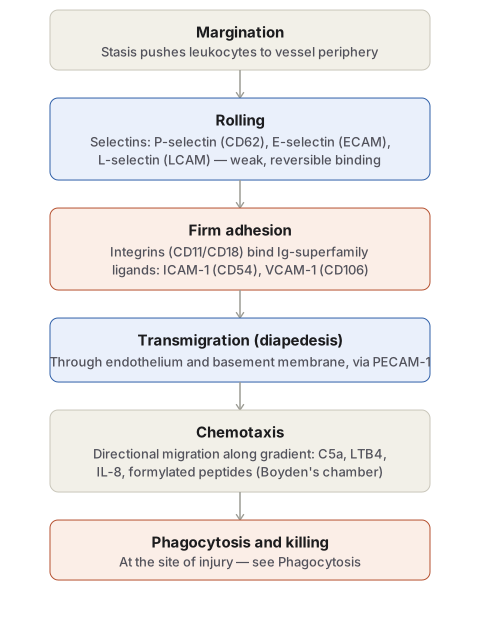
Margination → rolling (selectins: P-CD62, E-ECAM, L-LCAM) → firm adhesion (integrins CD11/CD18 ↔ Ig-superfamily ICAM-1/VCAM-1) → transmigration (PECAM-1) → chemotaxis (C5a, LTB4, IL-8, formylated peptides; demonstrated via Boyden’s chamber) → phagocytosis (see Phagocytosis).
Morphologic patterns
- Exudate type: serous / fibrinous / purulent (suppurative) / haemorrhagic / catarrhal
- Pseudomembranous inflammation (diphtheria, irritant gases)
- Ulcer — acute (polymorphs) vs chronic (lymphocytes, plasma cells, fibrosis)
- Suppuration/abscess — pus = dead+living neutrophils + debris + fibrin; boil (furuncle) vs carbuncle (diabetics)
- Cellulitis — diffuse spread (hyaluronidase)
- Blood: bacteraemia (few, non-multiplying) < septicaemia (multiplying, toxaemia, DIC risk) < pyaemia (septic thrombi → pyaemic abscesses/septic infarcts)
Systemic effects
Fever (PGs, IL-1, TNF-α) · leucocytosis (bacterial→neutrophilia, viral→lymphocytosis, parasitic→eosinophilia; exception: typhoid→leucopenia) · lymphangitis/lymphadenitis · shock (TNF-α → vasodilatation, permeability, DIC risk).
Outcomes
Resolution (complete, e.g. lobar pneumonia) · healing (regeneration or fibrosis) · suppuration/abscess · chronic inflammation.
Clinical relevance
Cardinal signs map directly to mechanism (examiner asks for mechanism, not just the name). Exudate vs transudate at bedside = same Starling-force logic. Fate of inflammation (resolving vs abscess vs chronic) drives management (drainage needed once suppuration is established).
Definition
Inflammation is the complex local response of living vascularised tissue to injury, whether from infection or from tissue damage of any other cause. Its purpose is to deliver the cells and molecules of host defence — phagocytic leukocytes, antibodies and complement proteins — from the circulation to the site where they are needed, so that the offending agent can be eliminated and the tissue prepared for repair. Although inflammation is often experienced as harmful, it is fundamentally a protective response; without it, infections would go unchecked and wounds would never heal.
Inflammation is broadly divided into acute and chronic forms. Acute inflammation is the initial, rapid response, developing within minutes to hours and lasting from a few hours to a few days; its main features are the leakage of fluid and plasma proteins (oedema) and the emigration of leukocytes, predominantly neutrophils. When the acute response fails to clear the offending agent, it can progress to a more protracted, chronic phase in which tissue destruction and fibrosis proceed alongside continuing inflammation. The two are best contrasted directly:
| Feature | Acute inflammation | Chronic inflammation |
|---|---|---|
| Onset | Fast — minutes to hours | Slow — days |
| Cellular infiltrate | Mainly neutrophils | Monocytes/macrophages and lymphocytes |
| Tissue injury | Usually mild and self-limited | May be significant |
| Fibrosis | None | May be severe and progressive |
| Local and systemic signs | Prominent | Variable, usually modest |
Causes
- Infections — bacterial, viral, fungal and parasitic — and their toxins, recognised by the host through pattern-recognition mechanisms described below.
- Tissue necrosis from any cause — ischaemia (e.g. myocardial infarction), physical agents (mechanical trauma, thermal injury, radiation, electric shock, sudden changes in atmospheric pressure) or chemical injury (strong acids/alkalis, insecticides, herbicides). Necrotic cells release molecules that trigger inflammation even without infection, referred to as “sterile inflammation.”
- Foreign bodies such as sutures or talc, which also provoke sterile inflammation.
- Immune reactions (hypersensitivity) — autoimmune disease and allergy, in which an otherwise protective immune response is misdirected against self tissue or against harmless environmental substances.
Recognition of Microbes and Damaged Cells
Before an inflammatory reaction can begin, the offending agent must first be recognised. This recognition step is carried out by cellular receptors and circulating plasma proteins.
Cellular receptors. Tissue-resident sentinel cells — chiefly macrophages and dendritic cells — carry receptors that detect conserved molecular structures shared across whole classes of pathogens, called pathogen-associated molecular patterns (PAMPs): examples include bacterial lipopolysaccharide (endotoxin), peptidoglycans, mannose-rich oligosaccharides and unmethylated CpG DNA. Receptors that recognise these patterns are therefore called pattern-recognition receptors (PRRs); the best-characterised family are the Toll-like receptors (TLRs), single-pass transmembrane proteins present on the cell surface and in endosomes, so named after the founding Toll gene discovered in Drosophila. Activation of TLRs triggers production of cytokines that initiate inflammation.
A separate cytosolic sensing system recognises damage-associated molecular patterns (DAMPs) — molecules released or altered by cell damage itself, such as uric acid, ATP leaked from injured mitochondria, falling intracellular potassium, or DNA escaped into the cytoplasm. These cytosolic receptors, of the NOD-like receptor (NLR) family, assemble a multiprotein complex called the inflammasome, which activates interleukin-1 (IL-1) and thereby recruits leukocytes. The inflammasome is also implicated in inflammatory reactions to urate crystals (gout), cholesterol crystals (atherosclerosis) and amyloid deposits (Alzheimer disease); gain-of-function mutations in these cytosolic receptors cause rare autoinflammatory syndromes treatable with IL-1 antagonists.
Circulating proteins. Microbes that reach the bloodstream are recognised by plasma proteins including the complement system, mannose-binding lectin and collectins, which can destroy circulating organisms directly and, on reaching tissue, stimulate a local inflammatory reaction.
The overall sequence of an inflammatory reaction can be remembered as the five R’s: recognition of the offending agent, recruitment of blood cells and proteins to the tissue, removal of the agent, regulation of the reaction, and repair of the injured tissue.
Vascular Events (Haemodynamic Changes)
Acute inflammation begins with a stereotyped, sequential set of changes in local blood vessels, whose purpose is to slow the circulation at the site of injury and let plasma proteins and cells escape into the tissue.
- Transient vasoconstriction of arterioles, lasting only seconds, is the earliest and briefest response and may be absent altogether.
- Progressive vasodilatation, first of arterioles and then opening of previously closed capillary beds, follows and is the earliest sustained feature — it increases local blood flow (hyperaemia) and accounts for the redness (rubor) and heat (calor) of inflamed tissue. Histamine is the principal mediator, with prostaglandins, kinins and nitric oxide also contributing.
- Increased vascular permeability with exudation, chiefly at the level of postcapillary venules, follows from contraction of endothelial cells and consequent opening of interendothelial gaps; protein-rich fluid escapes into the extravascular tissue. This is the mechanism of tumor (swelling) among the cardinal signs.
- Slowing of the circulation and stasis result as fluid loss concentrates red cells within the narrowed vascular lumen, raising blood viscosity; stasis is seen histologically as vascular congestion and produces the visible localised redness.
- Margination of leukocytes follows directly from stasis, as neutrophils — no longer kept centrally by fast axial flow — move to occupy a peripheral position along the vessel wall, setting up the cellular events described next.
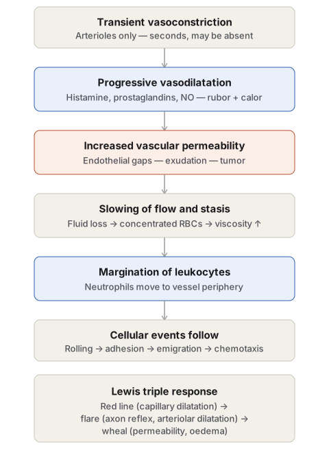
A related clinical demonstration of this sequence is the Lewis triple response, elicited by firmly stroking the skin: a red line appears within seconds from local capillary dilatation, followed by a spreading flare (arteriolar dilatation, an axon-mediated reflex) and finally a wheal, oedema from increased permeability at the stroked line itself.
Increased vascular permeability itself is produced through more than one mechanism, and recognising which mechanism is operating in a given setting is a common examination point:
| Mechanism | Timing | Example / mediators |
|---|---|---|
| Contraction of endothelial cells (formation of gaps) | Immediate, transient (15–30 min) | Histamine, bradykinin, leukotrienes, substance P — the commonest mechanism |
| Direct endothelial injury | Immediate, sustained until vessels thrombose or repair | Severe burns, infection |
| Delayed prolonged leakage | Onset delayed 2–12 hours, lasts days | Mild-to-moderate thermal injury, x-irradiation, ultraviolet injury, bacterial toxins |
| Leucocyte-mediated endothelial injury | At sites of leucocyte adhesion | Toxic oxygen species and proteolytic enzymes released by adherent neutrophils |
| Neovascularisation | Persists until endothelium matures | New capillaries in healing/repair remain leaky until endothelial junctions mature |
The fluid that escapes in inflammation, an exudate, is distinguished from a transudate — the fluid of oedema from purely haemodynamic imbalance, such as raised venous hydrostatic pressure or reduced plasma oncotic pressure — precisely because inflammatory exudation depends on increased vascular permeability rather than on Starling-force imbalance alone. Normally, capillary hydrostatic pressure (~32 mmHg at the arterial end, ~12 mmHg at the venous end) is balanced against a plasma colloid osmotic pressure of about 25 mmHg, so there is little net fluid movement; inflammation disrupts this balance by opening the vessel wall itself rather than by altering these pressures.
| Feature | Transudate | Exudate |
|---|---|---|
| Mechanism | Hydrostatic/osmotic imbalance, no permeability change | Increased vascular permeability |
| Protein content | Low | High |
| Specific gravity | Low | High |
| Cells | Few or none | Numerous (inflammatory cells) |
| Clinical setting | Congestive heart failure, hepatic or renal disease, protein malnutrition | Inflammation |
Cellular Events
The second major arm of acute inflammation is the recruitment of leukocytes — chiefly neutrophils — out of the vessel lumen and into the tissue, followed by their activation to destroy the offending agent.
Margination, rolling and adhesion. As blood flow slows, neutrophils marginate to the vessel periphery (above). They then bind loosely to endothelium and tumble along its surface — rolling — a process mediated by the selectins: P-selectin (CD62P, stored pre-formed in endothelial Weibel–Palade bodies and mobilised to the surface within minutes of histamine or thrombin exposure), E-selectin (ECAM, induced on cytokine-activated endothelium) and L-selectin (LCAM, expressed on leukocytes). Firm adhesion follows, mediated by leukocyte integrins (the CD11/CD18 family) binding to endothelial ligands of the immunoglobulin superfamily — ICAM-1 (CD54), VCAM-1 (CD106) and PECAM-1/CD31, the last of which also mediates the subsequent transmigration step.
Emigration (diapedesis). Firmly adherent leukocytes squeeze between endothelial cells, traverse the basement membrane, and move into the extravascular tissue, guided along the way by PECAM-1.
Chemotaxis. Once in the tissue, leukocytes migrate directionally along a concentration gradient toward the site of injury — a phenomenon classically demonstrated in the laboratory using Boyden’s chamber, in which leukocytes are shown to migrate toward, rather than merely more actively in the presence of, a chemoattractant. Chemotactic agents relevant to acute inflammation include bacterial products (formylated peptides), complement fragments C5a and C3a, leukotriene B4, interleukin-8, and, in the resolution phase, lipoxins, which act to inhibit further neutrophil recruitment (see Lipoxins).
Phagocytosis and killing. At the site of injury, neutrophils and macrophages recognise, engulf and destroy the offending agent through the recognition–engulfment–killing sequence described in full under Phagocytosis.
Morphologic Patterns of Acute Inflammation
Inflammation of an organ is conventionally named by adding the suffix -itis to its name (appendicitis, hepatitis, meningitis). Several recognisable morphologic patterns of acute inflammation are described, classified chiefly by the character of the exudate:
- Serous — a watery, low-cellularity exudate resembling serum, as in a tuberculous pleural effusion or a burn blister.
- Fibrinous — an exudate rich in fibrin, occurring where vascular leaks are large or there is a local procoagulant stimulus, as in pneumococcal or rheumatic pericarditis.
- Purulent (suppurative) — creamy pus formed in response to pyogenic bacteria, as in an abscess or acute appendicitis; discussed further below.
- Haemorrhagic — exudate containing red cells from vascular damage, as in the acute haemorrhagic pneumonia of influenza.
- Catarrhal — increased mucus secretion from surface epithelium, as in the common cold.
Pseudomembranous inflammation is a distinct pattern seen on mucosal surfaces (oral, respiratory, intestinal) exposed to diphtherial toxin or irritant gases: denuded epithelium allows plasma to exude onto the surface, where it coagulates together with necrotic epithelium to form a false membrane.
Ulcers are local surface defects produced by inflammation, typical of the stomach, duodenum, typhoid-affected intestine, intestinal tuberculosis, bacillary or amoebic dysentery, and varicose leg ulcers. Acute ulcers show polymorph infiltration with vasodilatation; long-standing ulcers instead show lymphocytes, plasma cells, macrophages and fibroblastic proliferation with scarring.
Suppuration (abscess formation) occurs when intense neutrophilic infiltration accompanying pyogenic bacterial infection causes tissue necrosis, forming a cavity filled with pus — a mixture of dead and living neutrophils, red cells, necrotic debris and fibrin, with macrophages and cholesterol crystals appearing as pus ages. Because of the tissue destruction involved, an abscess heals by fibrous scarring rather than by resolution. A boil (furuncle) is an abscess arising via a hair follicle; a carbuncle, typically in poorly controlled diabetics, is a loculated abscess spreading through the dermis and subcutaneous tissue of the neck.
Cellulitis is diffuse (non-localised) spread of inflammation through soft tissue, favoured by bacterial enzymes such as hyaluronidase that break down the tissue matrix.
Bacterial infection of the blood exists on a spectrum of severity: bacteraemia (small numbers of non-multiplying organisms, detected only by culture), septicaemia (rapidly multiplying, highly pathogenic organisms with systemic toxaemia, haemorrhages, neutrophilic leucocytosis and risk of disseminated intravascular coagulation), and pyaemia (dissemination of small septic thrombi that lodge and produce either multiple pyaemic abscesses, each with a necrotic bacteria-laden centre surrounded by suppuration, or larger septic infarcts when bigger thrombus fragments lodge in arteries).
Systemic Effects of Acute Inflammation
Beyond the local tissue response, acute inflammation produces recognisable systemic effects, together termed the acute phase response:
- Fever, driven by circulating pyrogens including prostaglandins, IL-1 and TNF-α released in response to infection.
- Leucocytosis, typically 15,000–20,000/µL; markedly higher counts with immature (“left-shifted”) myeloid forms constitute a leukaemoid reaction. Bacterial infection classically produces neutrophilia, viral infection lymphocytosis, and parasitic infestation eosinophilia — though typhoid fever is a notable exception, producing leucopenia with relative lymphocytosis despite being an acute bacterial infection.
- Lymphangitis and lymphadenitis, reactive inflammatory change in the lymphatics and nodes draining the inflamed site, with follicular hyperplasia and sinus histiocytosis on histology.
- Shock, in severe cases, driven by massive systemic release of TNF-α causing profuse vasodilatation, increased vascular permeability and intravascular volume loss, with the potential to progress to disseminated intravascular coagulation and death.
Outcomes (Fate) of Acute Inflammation
An episode of acute inflammation can end in one of four ways, depending chiefly on the extent of tissue destruction and whether the offending agent is successfully cleared:
- Resolution — complete return to normal structure and function, when tissue change has been slight and cellular injury reversible, as typically follows lobar pneumonia.
- Healing — by regeneration when tissue loss is superficial and the affected cells are capable of proliferation, or by fibrosis (scarring) when tissue destruction has been extensive.
- Suppuration — progression to abscess formation when pyogenic organisms have caused substantial tissue necrosis, as described above; an undrained abscess may eventually organise with dense fibrous tissue and calcify.
- Chronic inflammation — when the acute process persists or recurs, inflammation and healing then proceed side by side over a protracted course.
Clinical Relevance
- The cardinal signs are not arbitrary descriptors but a direct readout of the underlying vascular sequence — redness and heat from vasodilatation, swelling from increased permeability, pain from mediators such as prostaglandins and bradykinin acting on nerve endings, and loss of function from the combined effect of pain, swelling and tissue damage — which is why examiners repeatedly ask for the sign to be explained in terms of its mechanism, not simply named.
- Recognising which mechanism of increased vascular permeability is in play (immediate transient endothelial-gap formation versus direct endothelial injury versus delayed prolonged leakage) has direct clinical relevance, since it explains why a mild sunburn leaks for hours after the causative exposure has ended while a histamine-mediated wheal in a healthy person subsides within half an hour.
- Distinguishing an exudate from a transudate at the bedside — by protein content, specific gravity and cellularity of a pleural, peritoneal or pericardial tap — is a direct clinical application of the same Starling-force reasoning used to explain vascular permeability, and is the everyday way this topic is tested in the clinics.
- Correctly identifying the fate an episode of inflammation is heading toward (resolution versus suppuration versus chronicity) determines management — an early collection managed expectantly may still resolve, whereas an established abscess requires drainage because tissue destruction has already made resolution impossible.
Diagram 1: Vascular Events of Acute Inflammation
Draw a single downward column of five stages, each a labelled box connected to the next by a straight arrow.
- Transient vasoconstriction — arterioles only, lasts seconds, may be absent altogether.
- Progressive vasodilatation — mediators: histamine, prostaglandins, nitric oxide; produces redness and heat.
- Increased vascular permeability — endothelial gaps open, exudation begins; produces swelling.
- Slowing of flow and stasis — RBCs concentrate, viscosity rises.
- Margination of leukocytes — sets up the cellular events.
Below the fifth box, add a short note box stating that cellular events (rolling → adhesion → emigration → chemotaxis) follow directly, and a final box summarising the Lewis triple response (red line → flare → wheal) as the clinical demonstration of this same sequence.
Labels required
- Each stage must be numbered in sequence — examiners specifically test whether the order is known, not just the list of changes.
- The mediators driving vasodilatation and permeability (histamine in particular) must be named at their respective steps.
Errors commonly made
- Reversing the order of vasodilatation and increased permeability, or treating them as simultaneous rather than sequential.
- Omitting the transient vasoconstriction step altogether, since it is brief and easy to forget, even though it is frequently asked as “what is the earliest change.”
Diagram 2: Leukocyte Recruitment Cascade
Draw a single downward column of five stages, each box naming both the step and its molecular mediator(s).
- Margination — no named mediator, purely a consequence of stasis.
- Rolling — selectins (P-, E-, L-selectin).
- Firm adhesion — integrins (CD11/CD18) binding Ig-superfamily ligands (ICAM-1, VCAM-1).
- Transmigration — PECAM-1 (CD31).
- Chemotaxis — named chemoattractants (C5a, LTB4, IL-8, formylated bacterial peptides).
A final box for phagocytosis and killing closes the sequence, with a note directing to the Phagocytosis topic for the detailed mechanism.
Labels required
- Every step from rolling onward must carry its specific mediator family by name — “adhesion molecules” alone, without naming which family does which job, is the most common way marks are lost on this topic.
- Rolling (selectins, weak/reversible) must be visually or verbally distinguished from firm adhesion (integrins, strong/stable) since examiners frequently ask which molecule family is responsible for which step.
Errors commonly made
- Conflating selectins and integrins, or assigning ICAM-1/VCAM-1 to the rolling step instead of firm adhesion.
- Omitting PECAM-1’s specific role in the transmigration step and describing it only as “the cell squeezes through.”
Long Essays (2)
Short Essays (2)
Multiple Choice Questions (5)
My Sticky Notes
Personal revision notes, mnemonics and reminders.
