
Bacteriophage
Basics
Virus infecting bacteria. Described by Twort and d’Herelle (1917). Tadpole-shaped, 28-100 nm. Hexagonal head (capsid, dsDNA) + tail + tail fibers.
Lytic (Virulent) Cycle
Adsorption: random collision, tail fibers bind specific cell wall receptors.
Penetration: phage = syringe. Lysozyme (tail tip) makes hole in cell wall → DNA injected. Capsid stays outside as “ghost.” No uncoating step (nothing but DNA entered).
Biosynthesis: DNA + capsid proteins made.
Maturation/Assembly: DNA + head + tail proteins → daughter virions.
Release: phage enzymes lyse cell → daughter phages released.
Eclipse phase: DNA entry → first infectious phage. 15-30 min (much shorter than animal virus 15-30 hr).
Lysogenic (Temperate) Cycle
Phage DNA integrates into chromosome = prophage. Host = lysogenic bacterium, unharmed.
Lysogeny: prophage replicates synchronously with host chromosome, like a normal segment.
Lysogenic conversion: prophage confers new properties (toxin genes) on host.
- C. diphtheriae — diphtheria toxin from lysogenic phage. Cured phage = nontoxigenic.
- Other phage-coded toxins: Cholera toxin, Shiga toxin, E. coli verocytotoxin, Botulinum toxin C & D, Streptococcal pyrogenic exotoxin A & C.
Lysogenic→lytic interconversion: prophage excises → becomes lytic phage → replicates in cytoplasm → lyses cell.
Clinical/Practical Significance
Transduction: phage accidentally packages host DNA, transfers to next bacterium. Route for spreading plasmid-coded resistance (e.g. penicillin resistance in staph).
Phage typing: standardized phage panel subtypes bacteria below species level (classic for S. aureus). Largely replaced by molecular typing now.
Phage therapy: lytic phages used to treat infection (post-burn, wound infections). Renewed interest due to AMR concern.
Cloning vector: one of 4 major vector types (with plasmids, cosmids, artificial chromosomes) in recombinant DNA tech.
Codes for toxins: many major bacterial exotoxins are actually phage genes (see lysogenic conversion list above).
What a Bacteriophage Is
A bacteriophage — often shortened to “phage” — is simply a virus whose host is a bacterium rather than an animal cell, first described by Twort and d’Herelle in 1917. In structure it is unmistakable: a hexagonal protein head (capsid) enclosing double-stranded DNA, connected to a tail that ends in tail fibers, giving the whole particle a characteristic tadpole shape roughly 28–100 nm in size.
Two Life Cycles
Like the temperate bacteria-infecting viruses described more generally elsewhere, phages can pursue either a destructive or a quiescent strategy inside their host, and the choice has real downstream consequences for how genes move between bacteria.
The Lytic (Virulent) Cycle
This is the straightforward destructive pathway, followed by virulent phages, and it runs through the same broad stages as any other virus’s replication:
- Adsorption — the phage collides randomly with a bacterial cell and its tail fibers attach to specific receptors on the bacterial cell wall.
- Penetration — the phage behaves like a syringe: lysozyme carried at the tip of the tail punches a hole in the bacterial cell wall, and the phage DNA alone is injected through it into the cytoplasm. The empty capsid remains outside, still attached to the wall, as a so-called “ghost” — and because nothing but naked DNA actually entered the cell, there is no uncoating step at all, unlike the replication cycle of viruses that infect eukaryotic cells.
- Biosynthesis — the phage DNA and capsid proteins are synthesized using the hijacked machinery of the host.
- Maturation and assembly — DNA, head, and tail proteins are assembled into complete, infective daughter virions.
- Release — phage-coded enzymes lyse the bacterial cell, releasing the daughter phages to infect fresh bacteria.
The eclipse phase — the interval between DNA entry and the appearance of the first infectious phage particle, during which nothing can be detected inside the cell — lasts about 15–30 minutes, considerably shorter than the analogous phase in animal virus infections (15–30 hours), reflecting the phage’s much simpler replication needs.
The Lysogenic (Temperate) Cycle
Temperate phages instead integrate their DNA into the bacterial chromosome, where it is called a prophage, and the host bacterium is left unharmed. A bacterium carrying a prophage is a lysogenic bacterium, and the prophage behaves exactly like an ordinary segment of the host chromosome — replicating synchronously with it at every division, a phenomenon called lysogeny.
Two further things can happen from this quiescent state:
- Lysogenic conversion — while integrated, the prophage can confer entirely new properties on its host, most importantly the ability to make a toxin. The defining example is Corynebacterium diphtheriae: diphtheria toxin is encoded by a lysogenic phage, and a strain cured of that phage becomes nontoxigenic. Other phage-coded toxins follow the same logic — cholera toxin, shiga toxin, E. coli verocytotoxin, botulinum toxins C and D, and streptococcal pyrogenic exotoxins A and C.
- Lysogenic-to-lytic interconversion — a prophage is not necessarily permanent. When it eventually excises from the chromosome, it reverts to behaving as a lytic phage, replicating in the cytoplasm and ultimately lysing the cell to release progeny — the same destructive endpoint the lytic cycle reaches directly.
Why Bacteriophages Matter Beyond Their Own Biology
Phages are not just a curiosity of bacterial virology — they are a working tool and a mechanism of bacterial evolution in their own right:
- Transduction — a phage can accidentally package a fragment of host bacterial DNA instead of (or alongside) its own genome during a lytic infection, and carry that DNA into the next bacterium it infects. This is a genuine route for spreading plasmid-coded antibiotic resistance between bacteria — for instance, plasmid-coded penicillin resistance in staphylococci moves this way.
- Phage typing — because a given phage’s tail fibers recognize very specific bacterial surface receptors, a standardized panel of phages that will or will not infect a given strain can be used to subtype a bacterial species below the species level (classically used for S. aureus). Molecular typing methods have largely superseded this now, but it remains conceptually important.
- Phage therapy — because lytic phages kill bacteria directly, they have been explored as a treatment for bacterial infection, particularly for difficult wound and post-burn infections, and interest in this approach has grown again alongside concern about antimicrobial resistance.
- Cloning vector — phages are one of the standard vector classes (alongside plasmids, cosmids, and artificial chromosomes) used to carry foreign DNA in recombinant DNA technology.
- Coding for toxins — as already described under lysogenic conversion, a substantial share of the best-known bacterial exotoxins are not, strictly speaking, bacterial genes at all — they are phage genes, carried and expressed courtesy of a lysogenic infection.
Diagram 1: Lytic vs Lysogenic Cycle
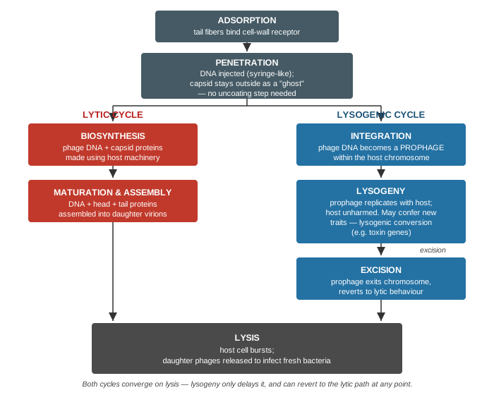
What to draw: A shared entry sequence (adsorption → penetration) branching into two paths — lytic (biosynthesis → assembly) and lysogenic (integration as prophage → lysogeny → excision) — with both paths converging on a shared final lysis step.
Common exam-marking mistakes to avoid:
- Do not draw an uncoating step for the phage — unlike animal viruses, only naked DNA enters the cell during penetration, so there is nothing to uncoat. This is one of the most commonly tested points of contrast between phage and animal-virus replication.
- Do not present the lysogenic cycle as a dead end — it is not an alternative to lysis, only a delay; the prophage can excise and revert to the lytic pathway at any point, and the diagram should show this explicitly rather than implying lysogeny is permanent.
- Label lysogenic conversion as a property of the lysogeny step specifically (the prophage sitting integrated), not of excision or lysis — the new trait (e.g. diphtheria toxin production) is expressed while the prophage is quiescent and integrated, not after it leaves the chromosome.
My Sticky Notes
Personal revision notes, mnemonics and reminders.
